SUMMARY.
Reading edition
MAMMALIA.
Primates 4
Carnivora 2
Perissodactyla 16
Artiodactyla (?) 1
Amblypoda 3
Rodentia 1
— 27
AVES.
Incertæ sedis 4
— 4
REPTILIA.
Crocodilia 6
Chelonia 5
— 11
PISCES.
Teleocephali 3
Nematognathi 1
Cycloganoidei 6
Plagiostomata 3
Incertæ sedis 1
— 14
—
Total 56
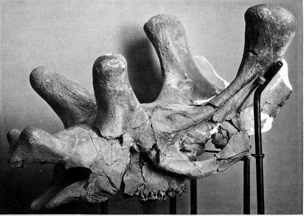
EXPLANATION OF PLATE A.
Figure about one fourth natural size.
Uintatherium Leidianum.
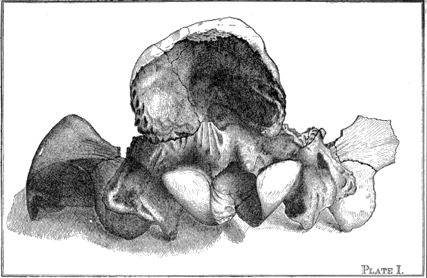
EXPLANATION OF PLATE I.
Figure three fourths natural size.
Palæosyops paludosus.
Posterior view of cranium.
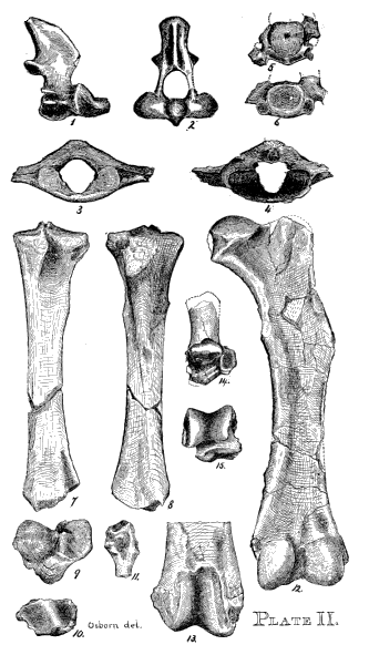
EXPLANATION OF PLATE II.
Figures one fourth natural size.
Palæosyops major.
Fig. 1.—Axis, view of right side.
Fig. 2.—Axis, anterior view.
Fig. 3.—Atlas, posterior view.
Fig. 4.—Alias, anterior view.
Figs. 5 and 6.—Seventh cervical anterior and posterior view
Figs. 7 and 8.—Anterior and posterior views of right tibia.
Figs. 9 and 10.—Proximal and distal faces of same.
Fig. 11.—Proximal end of fibula.
Fig. 12.—Right femur, posterior view.
Fig. 13.—Trochlea of the same.
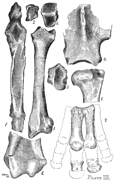
EXPLANATION OF PLATE III.
Figures one half natural size.
Palæosyops paludosus.
Figs. 1 and 2.—Ulna, anterior view, and distal articular face of same.
Figs. 3, 4, and 5.—Radius, anterior view; and distal and proximal faces of same.
Fig. 6.—Left scapula.
Fig. 7.—Right humerus, proximal end, anterior view.
Fig. 9.—Left manus, anterior view.
Palæosyops major.
Fig. 8.—Left humerus, anterior view.
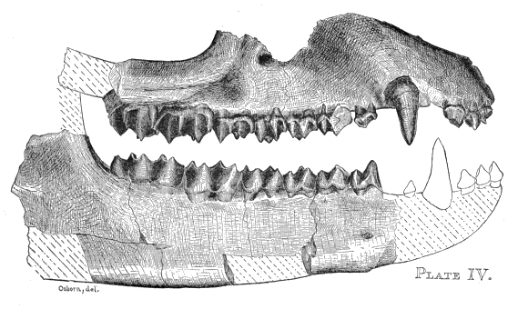
EXPLANATION OF PLATE IV.
Figure one half natural size.
Leurocephalus cultridens.
Upper and lower jaws. View of right side. Dotted outlines indicate probable position of lower incisor and canine series.
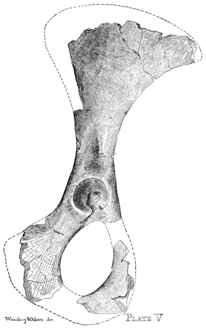
EXPLANATION OF PLATE V.
Figure one half natural size.
Palæosyops major.
Right innominate bone. The iliac crest and position of pubis estimated in outline.
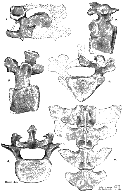
EXPLANATION OF PLATE VI.
Figures one third natural size.
Uintatherium leidianum.
Fig. 1.—Cervical vertebra, fifth or sixth, anterior view.
Fig. 2.—Dorsal vertebra, anterior region, view of left side.
Fig. 3.—Dorsal vertebra, middle region, posterior view.
Fig. 4.—Last lumbar vertebra, view of right side.
Fig. 5.—Last lumbar vertebra, posterior view.
Fig. 6.—Caudal series, first four vertebræ.
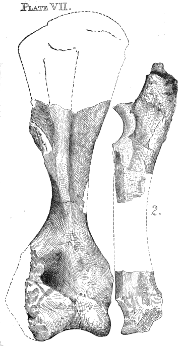
EXPLANATION OF PLATE VII.
Figures one third natural size.
Uintatherium leidianum.
Fig. 1.—Anterior view of right humerus.
Fig. 2.—Ulna, side view, length estimated.
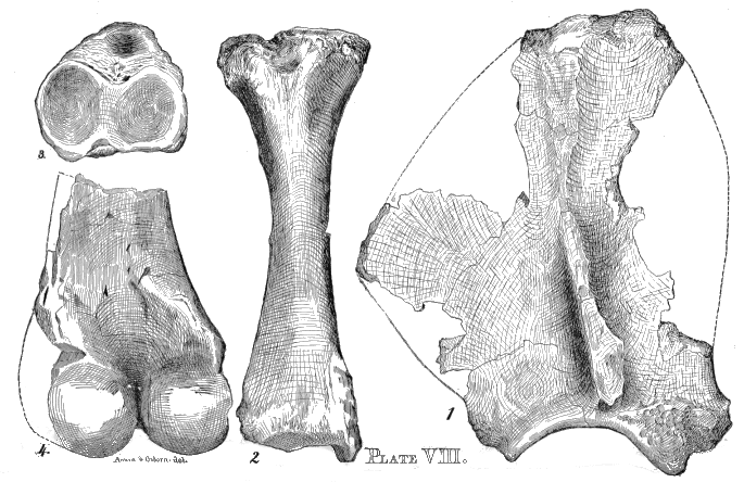
EXPLANATION OF PLATE VIII.
Figures one third natural size.
Uintatherium leidianum.
Fig. 1.—-Right scapula. The dotted outline gives an attempted restoration of the original shape.
Fig. 2.—Tibia of left side, anterior view.
Fig. 3.—View of the proximal articular faces of same.
Fig. 4.—Right femur, posterior view.
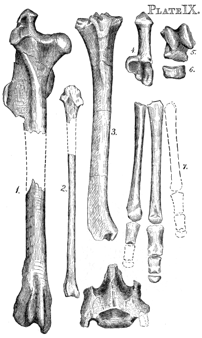
EXPLANATION OF PLATE IX.
Figures four fifths natural size.
Orohippus major.
Fig. 1.—Right femur, anterior view, length estimated.
Fig. 2.—Right fibula.
Fig. 3.—Right tibia, anterior view.
Fig. 4.—Calcaneum.
Figs. 5 and 6.—Astragalus and navicular.
Fig. 7.—Metatarsals and phalanges.
Last Fig.—A carnivorous sacrum, probably belonging to the brain, described on pp. 20-22.
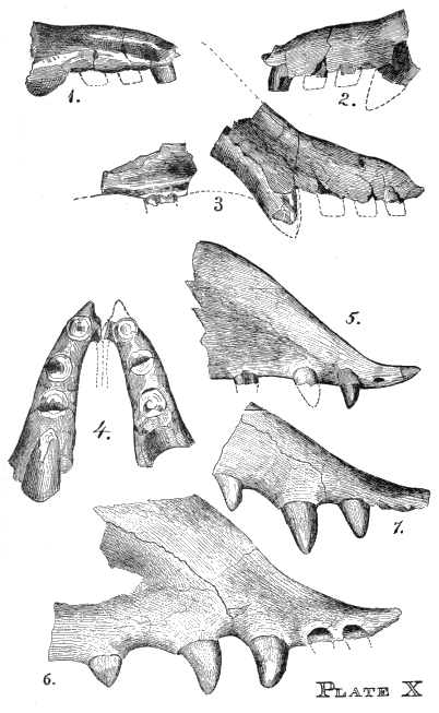
EXPLANATION OF PLATE X.
Figures 1, 2, 3, 4, and 6, natural size.
Ithygrammodon cameloides.
Fig. 1.—Left premaxillary, inner view.
Fig. 2.—Left premaxillary, outer view.
Fig. 3.—Right maxillary and premaxillary, outer view, premolar, with dotted outline indicating its probable position.
Fig. 4.—The premaxillaries in position (probable), view from above, showing size and position of the incisors. Dotted lines indicate the premaxillary spine (estimated).
Procamelus occidentalis, Cope, (See Wheeler's Survey, vol. iv., Plate LXXVII.)
Fig. 5.—Part of right maxillary and premaxillary, showing single incisor with rudimentary alveolus for a second.
Protolabis ——? Cope. (See Wheeler's Survey, vol iv., p. 343.)
Fig. 6.—Right maxillary and premaxillary. (This has never been drawn before, and was kindly lent to us for this purpose by Prof. Cope.)
Camelus bactrianus. Modern camel.
Fig. 7.—Right maxillaries, one half natural size.