Chapter II
Reading edition
THE INFLUENCE OF EXTERNAL CONDITIONS ON THE FORM AND STRUCTURE OF LARVÆ.
The facts recapitulated briefly in the preceding chapter show, that the forms of insect larvæ depend greatly on the group to which they belong. Thus the same tree may harbour larvæ of Diptera, Hymenoptera, Coleoptera, and Lepidoptera; each presenting the form typical of the family to which it belongs.
If, again, we take a group, such, for instance, as the Lamellicorn beetles, we shall find larvæ extremely similar in form, yet very different in habits. Those, for instance, of the common cockchafer (Fig. 1) feed on the roots of grass; those of Cetonia aurata (Fig. 2) inhabit ants' nests; the larvæ of the genus Trox (Fig. 3) are found on dry animal substances; of Oryctes (Fig. 4) in tan-pits; of Aphodius (Fig. 5) in dung; of Lucanus (the stag-beetle, Fig. 6) in wood.
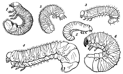
Fig. 1, Larva of the Cockchafer (Melolontha). (Westwood, Int. to the Modern Classification of Insects, vol. i. p. 194.). 2, Larva of Cetonia. 3, Larva of Trox. 4, Larva of Oryctes. 5, Larva of Aphodius (Chapuis and Candèze, Mém. Soc. Roy. Liège, 1853). 6, Larva of Lucanus. (Packard, Guide to the Study of Insects, Fig. 403).
On the other hand, in the present chapter it will be my object to show that the form of the larva depends very much on the conditions of its life. Thus, those larvæ which are internal parasites, whether in animals or plants, are vermiform, as are those which live in cells, and depend on their parents for food. On the other hand, larvæ which burrow in wood have strong jaws and generally somewhat weak thoracic legs; whilst those which feed on leaves have the thoracic legs more developed, but less so than the carnivorous species. Now, the Hymenoptera, as a general rule, belong to the first category: the larvæ of the Ichneumons, &c., which live in animals,—those of the Cynipidæ, inhabiting galls,—and those of ants, bees, wasps, &c., which are fed by their parents, are fleshy, apodal grubs; though the remarkable fact that the embryos of bees in one stage of their development possess rudiments of thoracic legs which subsequently disappear, seems to show, not indeed that the larvæ of bees were ever hexapod, but that bees are descended from ancestors which had hexapod larvæ, and that the present apod condition of these larvæ is not original, but results from their mode of life.
On the other hand, the larvæ of Sirex (Fig. 14) being wood-burrowers, possess well-developed thoracic legs. Again, the larvæ of the Tenthredinidæ, which feed upon leaves, closely resemble the caterpillars of Lepidoptera, even to the presence of abdominal pro-legs.
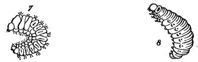
Fig. 7, Larva of Brachytarsus (Ratzeburg, Forst. Insecten). 8, Larva of Crioceris (Westwood, loc. cit.).
The larvæ of most Coleoptera (Beetles) are active, hexapod, and more or less flattened: but those which live inside vegetable tissues, such as the weevils, are apod fleshy grubs, like those of Hymenoptera. Pl. II., Fig. 6, represents the larva of the nut-weevil, Balaninus (Pl. I., Fig. 6), and it will be seen that it closely resembles Pl. II., Fig. 5, which represents that of a fly (Anthrax), Pl. I., Fig. 5, and Pl. II., Figs. 7, 8, and 9, which represent respectively those of a Cynips or gall-fly (Pl. I., Fig. 7), an ant (Pl. I., Fig. 8), and wasp (Pl. I., Fig. 9). Nor is Balaninus the only genus of Coleoptera which affords us examples of this fact. Thus in the genus Scolytus (Pl. I., Fig. 4), the larvæ (Pl. II., Fig. 4), which, as already mentioned, feed on the bark of the elm, closely resemble those just described, as also do those of Brachytarsus (Fig. 7). On the other hand, the larvæ of certain beetles feed on leaves, like the caterpillars of Lepidoptera; thus that of Crioceris Asparagi (Fig. 8)—which, as its name denotes, feeds on the asparagus—closely resembles the larvæ of certain Lepidoptera, as for instance of Thecla spini. From this point of view the transformations of the genus Sitaris (Pl. III., Fig. 4), which have been very carefully investigated by M. Fabre, are peculiarly interesting.12
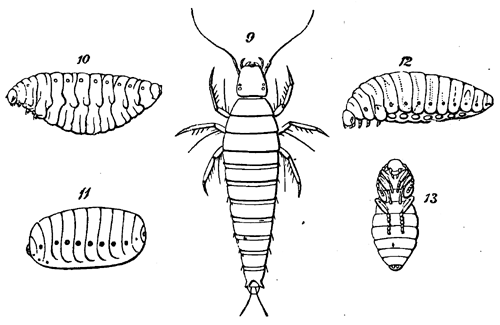
Fig. 9, Larva of Sitaris humeralis (Fabre, Ann. des Sci. Nat., sér. 4, tome vii.). 10, Larva of Sitaris humeralis, in the second stage. 11, Larva of Sitaris humeralis, in the third stage. 12, Larva of Sitaris humeralis, in the fourth stage. 13, Pupa of Sitaris.
The genus Sitaris (a small beetle allied to Cantharis, the blister-fly, and to Meloë, the oil-beetle) is parasitic on a kind of Bee (Anthophora), which excavates subterranean galleries, each leading to a cell. The eggs of the Sitaris, which are deposited at the entrance of these galleries, are hatched at the end of September or beginning of October; and M. Fabre not unnaturally expected that the young larvæ, which are active little creatures with six serviceable legs (Fig. 9), would at once eat their way into the cells of the Anthophora. No such thing: till the month of April following they remain without leaving their birthplace, and consequently without food; nor do they in this long time change either in form or size. M. Fabre ascertained this, not only by examining the burrows of the Anthophoras, but also by direct observation of some young larvæ kept in captivity. In April, however, his captives at last awoke from their long lethargy, and hurried anxiously about their prisons. Naturally inferring that they were in search of food, M. Fabre supposed that this would consist either of the larvæ or pupæ of the Anthophora, or of the honey with which it stores its cell. All three were tried without success. The first two were neglected, and the larvæ, when placed on the latter, either hurried away, or perished in the attempt, being evidently unable to deal with the sticky substance. M. Fabre was in despair: “Jamais expérience,” he says, “n’a éprouvé pareille déconfiture. Larves, nymphes, cellules, miel, je vous ai tous offert; que voulez-vous donc, bestioles maudites?” The first ray of light came to him from our countryman, Newport, who ascertained that a small parasite found by Léon Dufour on one of the wild bees, and named by him Triungulinus, was, in fact, the larva of Meloë;. The larvæ of Sitaris much resembled Dufour’s Triungulinus; and acting on this hint, M. Fabre examined many specimens of Anthophora, and found on them at last the larvæ of his Sitaris. The males of Anthophora emerge from the pupæ sooner than the females, and M. Fabre ascertained that, as they come out of their galleries, the little Sitaris larvæ fasten upon them. Not, however, for long: instinct teaches them that they are not yet in the straight path of development; and, watching their opportunity, they pass from the male to the female bee. Guided by these indications, M. Fabre examined several cells of the Anthophora: in some, the egg of the Anthophora floated by itself on the surface of the honey; in others, on the egg, as on a raft, sat the still more minute larva of the Sitaris. The mystery was solved. At the moment when the egg is laid the Sitaris larva springs upon it. Even while the poor mother is carefully fastening up her cell, her mortal enemy is beginning to devour her offspring: for the egg of the Anthophora serves not only as a raft, but as a repast. The honey which is enough for either, would be too little for both; and the Sitaris, therefore, at its first meal, relieves itself from its only rival. After eight days the egg is consumed, and on the empty shell the Sitaris undergoes its first transformation, and makes its appearance in a very different form, as shown in Fig. 10.
The honey which was fatal before is now necessary; the activity which before was necessary is now useless; consequently, with the change of skin, the active, slim larva changes into a white, fleshy grub, so organized as to float on the surface of the honey, with the mouth beneath, and the spiracles above the surface: “grâce à l’embonpoint du ventre,” says M. Fabre, “la larve est à l’abri de l’asphyxie.” In this state it remains until the honey is consumed; then the animal contracts, and detaches itself from its skin, within which the further transformations take place. In the next stage, which M. Fabre calls the pseudo-chrysalis (Fig. 11), the larva has a solid corneous envelope and an oval shape; and in its colour, consistency, and immobility reminds one of a Dipterous pupa. The time passed in this condition varies much. When it has elapsed, the animal moults again, again changes its form, and assumes that shown in Fig. 12; after this it becomes a pupa (Fig. 13) without any remarkable peculiarities. Finally, after these wonderful changes and adventures, in the month of August the perfect Sitaris (Pl. III., Fig. 4) makes its appearance.
On the other hand, there are cases in which larvæ diverge remarkably from the ordinary type of the group to which they belong, without, as it seems in our present imperfect state of information, any sufficient reason.
Thus the ordinary type of Hymenopterous larva, as we have already seen, is a fleshy apod grub; although those of the leaf-eating and wood-boring groups, Tenthredinidæ and Siricidæ (Fig. 14), are caterpillars, more or less closely resembling those of Lepidoptera. There is, however, a group of minute Hymenoptera, the larvæ of which reside within the eggs or larvæ of other insects. It is difficult to understand why these larvæ should differ from those of Ichneumons, which are also parasitic Hymenoptera, and should be, as will be seen by the accompanying figures, of such remarkable and grotesque forms. The first known of these curious larvæ was observed by De Filippi,13 who, having collected some of the transparent eggs of a small Beetle (Rhynchites betuleti), to his great surprise found more than half of them attacked by a parasite, which proved to be the larva of a minute Hymenopterous insect belonging to the Pteromalidæ. Fig. 15 shows the egg of the Beetle, with the parasitic larva, which is represented on a larger scale in Fig. 16.
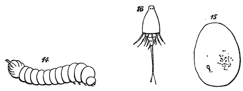
Fig. 14, Larva of Sirex (Westwood, loc. cit.). 15, Egg of Rhynchites, showing the parasitic Larva in the interior. 16, the parasitic Larva more magnified.
More recently this group has been studied by M. Ganin,14 who thus describes the development of Platygaster. The egg, as in allied Hymenopterous families, for instance in Cynips, is elongated and club-shaped (Fig. 17). After a while a large nucleated cell appears in the centre (Fig. 18). This nucleated cell divides (Fig. 19) and subdivides. The outermost cells continue the same process, thus forming an outer investing layer. The central, on the contrary, enlarges considerably, and develops within itself a number of daughter cells (Figs. 20 and 21), which gradually form a mulberry-like mass, thus giving rise to the embryo (Fig. 22).
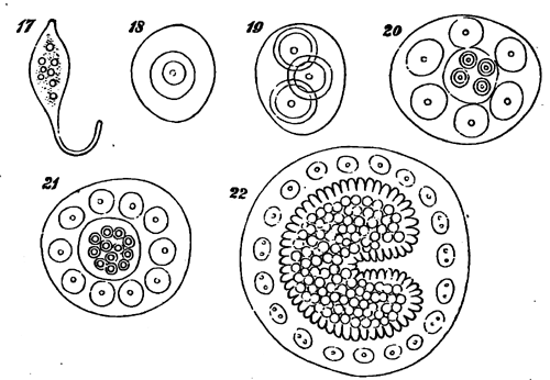
Fig. 17, Egg of Platygaster (after Ganin). 18, Egg of Platygaster showing the central cell. 19, Egg of Platygaster after the division of the central wall. 20, Egg of Platygaster more advanced. 21, Egg of Platygaster more advanced. 22, Egg of Platygaster showing the rudiment of the embryo.
Ganin met with the larvæ of Platygaster in those of a small gnat, Cecidomyia. Sometimes as many as fifteen parasites occurred in one gnat, but as a rule only one of these attained maturity. The three species of Platygaster differ considerably in form, as shown in Figs. 23-25. They creep about within the larva of Cecidomyia by means of the strong hooked feet, kf, somewhat aided by movements of the tail. They possess a mouth, stomach, and muscles, but the nervous, vascular, and respiratory systems do not make their appearance until later. After some time the larva (Fig. 23) changes its skin, assuming the form represented in Fig. 26. In this moult the last abdominal segment of the first larva is entirely thrown off: not merely the outer skin, as in the case of the other segments, but also the hypodermis and the muscles. This larva, as will be seen by the figure, resembles a barrel or egg in form, and is .870 mm. in length, the external appendages having disappeared, and the segments being indicated only by the arrangement of the muscles. slkf is the œsophagus leading into a wide stomach which occupies nearly the whole body, gsae is the rudiment of the supra-œsophageal ganglia, bsm the ventral nervous cords. The ventral nervous mass has the form of a broad band, with straight sides; it consists of embryonal cells, and remains in this undeveloped condition during the whole larval state.
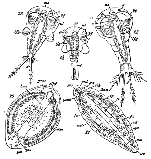
Fig. 23, Larva of Platygaster (after Ganin)—mo, mouth; a, antenna; kf, hooked feet; z, toothed process; lfg, lateral process; f, branches of the tail. 24, Larva of another species of Platygaster. The letters indicate the same parts as in the preceding figure. 25, Larva of a third species of Platygaster. The letters indicate the same parts as in the preceding figures. 26, Larva of Platygaster in the second stage—mo, mouth; slkf, œsophagus; gsae, supra-œsophageal ganglion; lm, muscles; bsm, nervous system; ga, gh, rudiments of the reproductive glands. 27, Larva of Platygaster in the third stage—mo, mouth; md, mandibles; gsae, supra-œsophageal ganglion; slk, œsophagus; ag, ducts of the salivary glands; bnm, ventral nervous system; sp, salivary glands; msl, stomach; im, imaginal discs; tr, tracheæ; fk, fatty tissue; ed, intestine; ga, rudiments of reproductive organs; ew, wider portion of intestine; ao, posterior opening.
At the next moult the larva enters its third state, which, as far as the external form (Fig. 27) is concerned, differs from the second only in being somewhat more elongated. The internal organs, however, are much more complex and complete. The tracheæ have made their appearance, and the mouth is provided with a pair of mandibles. From this point the metamorphoses of Platygaster do not appear to differ materially from those of other parasitic Hymenoptera.
An allied genus, Polynema, has also very curious larvæ. The perfect insect is aquatic in its habits, swimming by means of its wings; flying, if we may say so, under water.15 It lays its eggs inside those of Dragon-flies; and the embryo, as shown in Fig. 28, has the form of a bottle-shaped mass of undifferentiated embryonal cells, covered by a thin cuticle, but without any trace of further organization. Protected by the egg-shell of the Dragon-fly, and bathed in the nourishing fluid of the Dragon-fly’s egg, the young Polynema imbibes nourishment through its whole surface, and increases rapidly in size. The digestive canal gradually makes its appearance; the cellular mass forms a new skin beneath the original cuticle, distinctly divided into segments, and provided with certain appendages. After a while the old cuticle is thrown off, and the larva gradually assumes the form shown in Fig. 29. The subsequent metamorphoses of Polynema offer no special peculiarities.
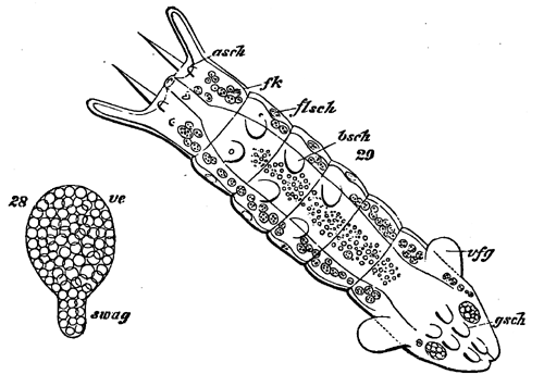
Fig. 28, Embryo of Polynema (after Ganin). 29, Larva of Polynema—asch, rudiments of the antenna; flsch, rudiments of the wings; bsch, rudiments of the legs; vfg. lateral projections; gsch, rudiments of the ovipositor; fk, fatty tissue.
From these facts—and, if necessary, many more of the same nature might have been brought forward—it seems to me evident that while the form of any given larva depends to a certain extent on the group of insects to which it belongs, it is also greatly influenced by the external conditions to which it is subjected; that it is a function of the life which the larva leads and of the group to which it belongs.
The larvæ of insects are generally regarded as being nothing more than immature states—as stages in the development of the egg into the imago; and this might more especially appear to be the case with those insects in which the larvæ offer a general resemblance in form and structure (excepting of course so far as relates to the wings) to the perfect insect. Nevertheless we see that this would be a very incomplete view of the case. The larva and pupa undergo changes which have no relation to the form which the insect will ultimately assume. With a general tendency to this goal, as regards size and the development of the wings, there are coincident other changes having reference only to existing wants and condition. Nor is there in this, I think, anything which need surprise us. External circumstances act on the insect in its preparatory states, as well as in its perfect condition. Those who believe that animals are susceptible of great, though gradual, change through the influence of external conditions, whether acting, as Mr. Darwin has suggested, through natural selection, or in any other manner, will see no reason why these changes should be confined to the mature animal. And it is evident that creatures which, like the majority of insects, live during the successive periods of their existence in very different circumstances, may undergo considerable changes in their larval organization, in consequence of forces acting on them while in that condition; not, indeed, without affecting, but certainly without affecting to any corresponding extent, their ultimate form.
I conclude, therefore, that the form of the larva in insects, whenever it departs from the hexapod Campodea type, has been modified by the conditions under which it lives. The external forces acting upon it are different from those which affect the mature form; and thus changes are produced in the young which have reference to its immediate wants, rather than to its final form.
And, lastly, as a consequence, that metamorphoses may be divided into two kinds, developmental and adaptional or adaptive.