PLATE XIV.
Reading edition
Fig 1.
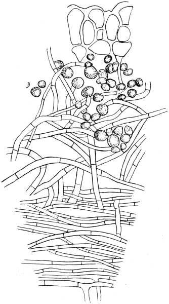
Fig 2.
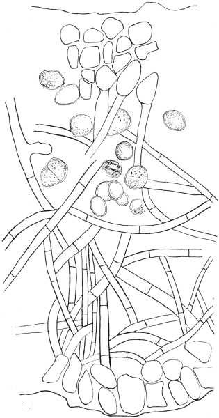
Fig 3.
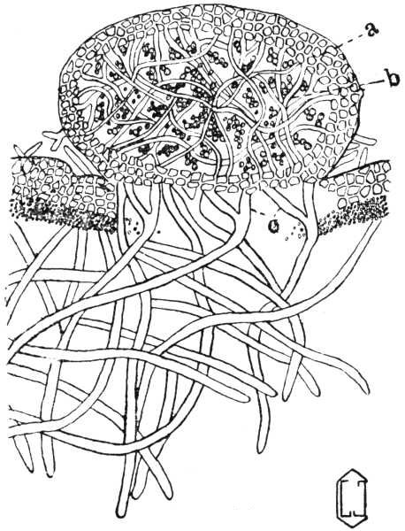
Fig 4.
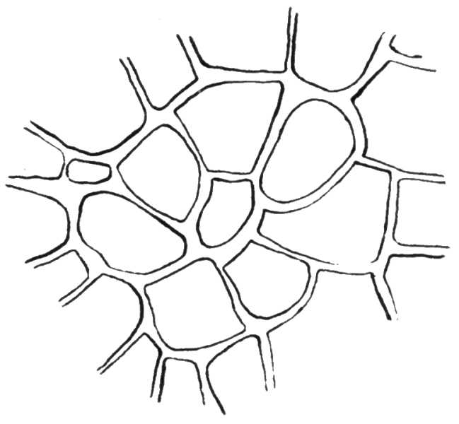
EXPLANATION OF PLATE XV
Fig. 5. A section through the cortex of Peltigera canina, showing its relation to the medullary hyphae. X 760.
Fig. 6. A portion of a section of the hymenium of Peltigera canina, showing two asci containing spores, two asci with protoplasmic contents, and five paraphyses. X 760.
Fig. 7. A portion of a section through an apothecium of Peltigera canina, showing part of the hymenium of interwoven hyphae below and the bases of three paraphyses above. X 760.
Fig. 8. Types of spores found in the Peltigeraceae: a, 4-celled spores of Peltigera horizontalis; b, 6- to 8-celled spores of Peltigera sorediata; c, 4-celled spores of Peltigera aphthosa; d, 4- to 8-celled spores of Peltigera canina. X 380.
The drawings were made with camera lucida and were reduced one-half in making the plates.