PLATE I.
Reading edition
ANATOMICAL.
1. Outline of a Lepidopterous insect showing the terms employed in describing the various margins and angles of the fore- and hind-wings. 2. View of the under side of the head and first segment of the larva of a Lepidopterous insect. AA, eyes; BB, antennæ; 1, labrum; 22, mandibles; 33, maxillæ; 4, labium; 5, spinneret; a, coxa; b, trochanter; c, femur; d, tibia; e, tarsus; f, claw (highly magnified). 3. Assumed type of neuration of fore-wing of a Lepidopterous insect. (After Meyrick.) 4. Ditto of hind-wing. (After Meyrick.) 5. Side view of the head of Vanessa gonerilla with proboscis extended. (Imago, Plate XII., fig. 5.) 6. Ditto with proboscis coiled up. (In both these figures only the basal portions of the antennæ are shown.) 7. Neuration of fore-wing of Anosia erippus. (Imago, Plate XI., fig. 1.) 8. Ditto of hind-wing. 9. Digestive system of a Lepidopterous larva. A, œsophagus; D, ventriculus; F, clavate intestine; E, ilium; H, colon; K, biliary vessels; O, spinning vessels. (After Suckow.) 10. Ditto of perfect insect. N, salivary vessels; C, sucking stomach; G, cæcum. The rest as before. (After Herold.) 11. Front view of the head of Vanessa gonerilla with the labial palpi removed showing the organs of the mouth. AA, eyes; BB, antennæ (basal portion); l, labrum; mm, mandibles; pp, maxillary palpi; C, proboscis formed of elongated maxillæ (highly magnified). 12. Neuration of fore-wing of Sphingidæ. (Deilephila; after Meyrick.) 13. Ditto hind-wing. (After Meyrick.) 14. Proleg of caterpillar highly magnified. 15. Neuration of fore-wing of Chrysophanus salustius. (Imago, Plate XII., figs. 18-21.) 16. Ditto of hind-wing. 17. Fasciculate-ciliated antenna of Chloroclystis plinthina. (Imago, Plate VI., fig. 8.) 18. Serrate antenna of Melanchra composita. (Imago, Plate V., fig. 8.) 19. Pubescent antenna of Epirranthis alectoraria. (Imago, Plate VIII., figs. 42-47.) 20. Bi-pectinated antenna of Nyctemera annulata. (Imago, Plate IV., figs. 1, 2.) 21. Leg of Agrotis ypsilon. (Imago, Plate V., figs. 35, 36.) 1, coxa; 2, trochanter; 3, femur; 4, tibia; 5, tarsus; 6, claw; SS, spurs. (All these are highly magnified.) 22. Neuration of fore-wing of Hepialus virescens. (Imago, Plate XIII., figs. 16, 17.) 23. Ditto of hind-wing. 24. Head of ditto. 25. Neuration of fore-wing of Erebia pluto. (Imago, Plate XI., figs. 8-10.) Vein 11 absent. 26. Ditto, veins 11 and 12 concurrent. 27. Ditto of hind-wing. 28. Neuration of fore-wing of Porina signata. (Imago, Plate XIII., fig. 15.) 29. Ditto of hind-wing. 30. Neuration of fore-wing of Œceticus omnivorus. (Imago, Plate XIII., fig. 6.) 31. Ditto of hind-wing.
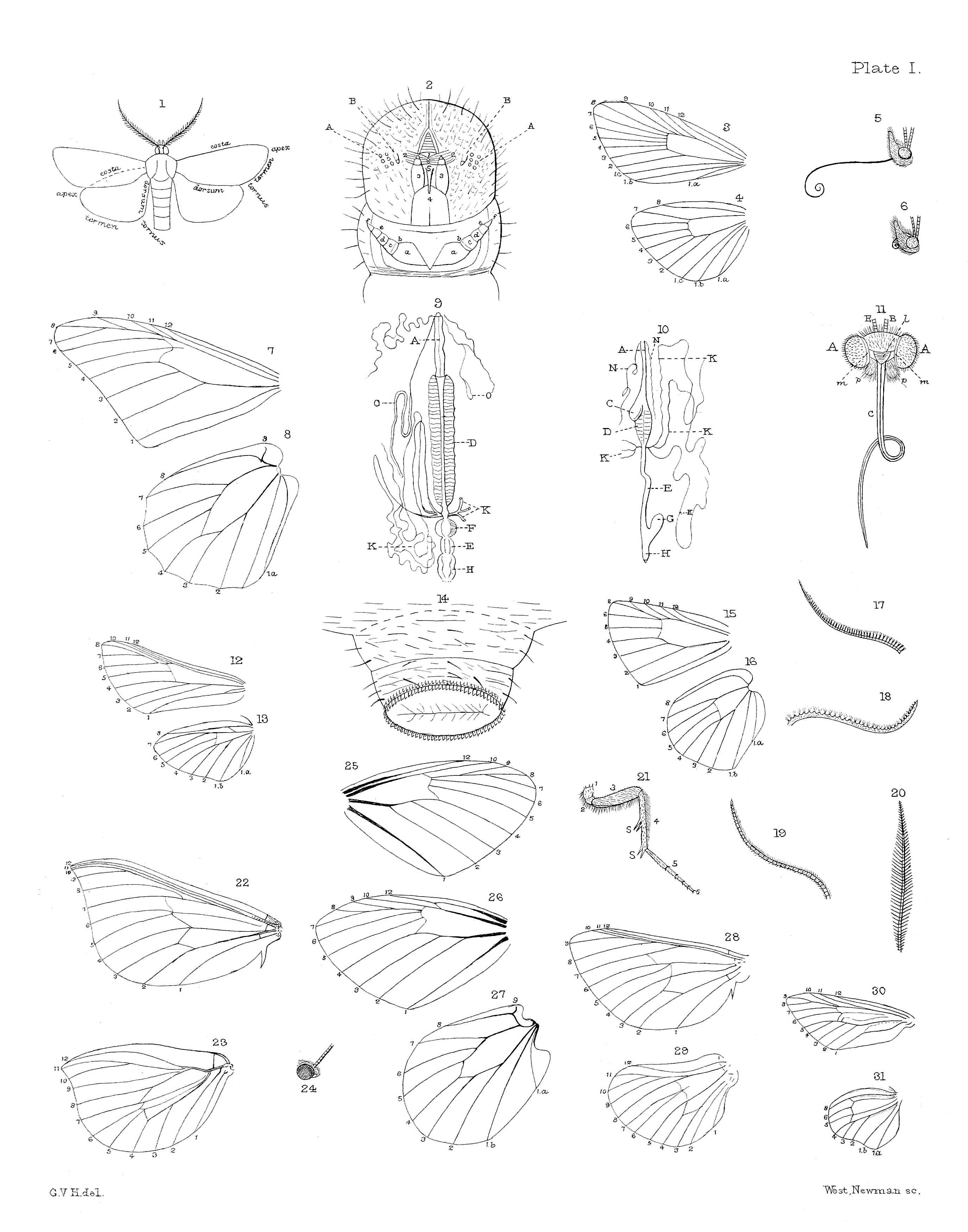
1. Outline of a Lepidopterous insect showing the terms employed in describing the various margins and angles of the fore- and hind-wings.
2. View of the under side of the head and first segment of the larva of a Lepidopterous insect. AA, eyes; BB, antennæ; 1, labrum; 22, mandibles; 33, maxillæ; 4, labium; 5, spinneret; a, coxa; b, trochanter; c, femur; d, tibia; e, tarsus; f, claw (highly magnified).
3. Assumed type of neuration of fore-wing of a Lepidopterous insect. (After Meyrick.)
4. Ditto of hind-wing. (After Meyrick.)
5. Side view of the head of Vanessa gonerilla with proboscis extended. (Imago, Plate XII., fig. 5.)
6. Ditto with proboscis coiled up. (In both these figures only the basal portions of the antennæ are shown.)
7. Neuration of fore-wing of Anosia erippus. (Imago, Plate XI., fig. 1.)
8. Ditto of hind-wing.
9. Digestive system of a Lepidopterous larva. A, œsophagus; D, ventriculus; F, clavate intestine; E, ilium; H, colon; K, biliary vessels; O, spinning vessels. (After Suckow.)
10. Ditto of perfect insect. N, salivary vessels; C, sucking stomach; G, cæcum. The rest as before. (After Herold.)
11. Front view of the head of Vanessa gonerilla with the labial palpi removed showing the organs of the mouth. AA, eyes; BB, antennæ (basal portion); l, labrum; mm, mandibles; pp, maxillary palpi; C, proboscis formed of elongated maxillæ (highly magnified).
12. Neuration of fore-wing of Sphingidæ. (Deilephila; after Meyrick.)
13. Ditto hind-wing. (After Meyrick.)
14. Proleg of caterpillar highly magnified.
15. Neuration of fore-wing of Chrysophanus salustius. (Imago, Plate XII., figs. 18-21.)
16. Ditto of hind-wing.
17. Fasciculate-ciliated antenna of Chloroclystis plinthina. (Imago, Plate VI., fig. 8.)
18. Serrate antenna of Melanchra composita. (Imago, Plate V., fig. 8.)
19. Pubescent antenna of Epirranthis alectoraria. (Imago, Plate VIII., figs. 42-47.)
20. Bi-pectinated antenna of Nyctemera annulata. (Imago, Plate IV., figs. 1, 2.)
21. Leg of Agrotis ypsilon. (Imago, Plate V., figs. 35, 36.) 1, coxa; 2, trochanter; 3, femur; 4, tibia; 5, tarsus; 6, claw; SS, spurs. (All these are highly magnified.)
22. Neuration of fore-wing of Hepialus virescens. (Imago, Plate XIII., figs. 16, 17.)
23. Ditto of hind-wing.
24. Head of ditto.
25. Neuration of fore-wing of Erebia pluto. (Imago, Plate XI., figs. 8-10.) Vein 11 absent.
26. Ditto, veins 11 and 12 concurrent.
27. Ditto of hind-wing.
28. Neuration of fore-wing of Porina signata. (Imago, Plate XIII., fig. 15.)
29. Ditto of hind-wing.
30. Neuration of fore-wing of Œceticus omnivorus. (Imago, Plate XIII., fig. 6.)
31. Ditto of hind-wing.