GARY N. CALKINS, (continued 2)
Reading edition
1. Body cylindrical; size medium; peristome long and turns sharply to the left at the extremity Genus Metopus
2. No torsion in the peristome; undulating membrane is confined to the posterior part of peristome Genus Blepharisma
3. No peristomial torsion; body highly contractile; no undulating membrane Genus Spirostomum
Key to the marine genera of Bursaridæ.
Diagnostic characters: The body is usually short and pocket-like, but may be elongate. The chief characteristic is the peristome, which is not a furrow, but a broad triangular area deeply insunk and ending in a point at the mouth. The adoral zone is usually confined to the left peristome edge, or it may cross over to the right anterior edge.
1. The anterior half of the body tapers to nearly a point in front; the peristome is narrowest at the apex; the mouth is the entire peristome base. Genus Balantidium
2. The anterior end does not taper; the peristome is widest at the end of the body; the mouth is clearly defined. Genus *Condylostoma
* Presence at Woods Hole indicated by asterisk.
Genus CONDYLOSTOMA (KONDYLOSTOMA Bory de St. Vincent 1824) Dujardin '41
(Dujardin '41; Claparède & Lachmann '58; Stein '59, '67; Cohn '66; Quennerstedt '67; Wrzesniowski '70; Bütschli '76, '88; Kent '81; Maupas '83; Shevyakov '96.)
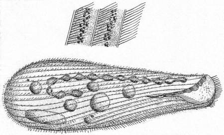
Colorless and more or less flexible animals of medium size. The general form is elongate and cylindrical or somewhat smaller anteriorly. The posterior end is broadly rounded, the anterior end somewhat truncate and oblique. The peristome is broad and triangular, the base of the triangle being the entire anterior end of the body. The entire length of the peristome is one-fourth or less of the body length. The mouth is large and placed at the apex of the peristomial triangle and opens into a comparatively small œsophagus. The right edge of the peristome is lamellate and bears a clearly defined undulating membrane. The adoral zone is well developed upon the left edge of the peristome, from which it passes around anteriorly to the right edge. The surface of the peristome is free from cilia, but the rest of the body is uniformly coated with small active cilia. Contractile vacuoles are not safely determined. Bütschli thinks there is probably one terminal vacuole, but some observers deny this (e.g. Maupas). Others describe them on the dorsal side of the posterior end (Quennerstedt). The macronucleus is long and beaded and placed upon the right side. Micronuclei are numerous and scattered along the macronucleus. The anus is terminal and dorsal. Food consists of large and small particles. Movement rapid, free swimming, alternating with resting periods; in some cases an undulating or wriggling movement is seen, showing clearly the flexibility of the body. Fresh and salt water.
Condylostoma patens Müller. Fig. 45.
The body is elongate, somewhat sac-like, five or six times as long as broad, plastic, and frequently contains brightly colored food granules. The triangular peristome takes up the greater part of the anterior end, and the mouth is situated at the sharper angle of the triangle, about one-fourth of the total length from the anterior end. The cuticle is longitudinally striated, the lines having a slightly spiral course. They are not closely set, and fine cilia are thickly inserted along their edges. The endoplasm is granular and viscous. The motile organs consist of an adoral zone of membranelles, which stretch along the left edge of the peristome and the front edge of the body. The right edge of the peristome supports an undulating membrane. The nucleus is moniliform and extends the full length of the left side; a number of micronuclei are distributed along its course (Maupas).
Length 400µ; diameter at widest part 105µ. Maupas gives the length from 305µ to 495µ; and Stein 376µ to 564µ. Very common.
For a more extended account of the structures, see the excellent description by Maupas '83.
Fig. 45.—Condylostoma patens. ENLARGE
Key to the marine genera of Stentoridæ.
Diagnostic characters: The peristome is relatively short and limited to the front end of the animal, so that its plane is nearly at right angles to that of the longitudinal axis of the body. The adoral zone of cilia either passes entirely around the peristome edge or ends at the right-hand edge. The surface of the peristome is spirally striated and provided with cilia. Undulating membranes are absent.
1. Peristome circular in outline; limited to the anterior end Genus Stentor
2. The peristome is drawn out into two wing-like processes; tube-dwelling Genus Folliculina
Genus STROMBIDIUM Cl. & Lach. '58.
(Stein '67; Bütschli '73; Fromentel '74; Kent '81; Gruber '84; Entz '84; Maupas '83. Bütschli '88.)
Small, colorless (except for ingested food) animals with characteristic springing movements. The form is usually constant, but in some cases may be plastic like Astasia; it is usually globular or conical, the posterior end being more or less pointed, the anterior end broadest. The latter is surrounded by a complete circle of the adoral zone, the oral end of which passes into a peristomial depression which extends deep into the middle of the body. The mouth, with a very small œsophagus, lies at the bottom of the inturned peristome. The region surrounded by the adoral zone is frequently drawn out into an anterior process, occasionally bearing a pigment mass. The ventral surface in some cases bears cilia, which may be distributed or restricted to a row of large cilia. Trichocysts are usually present and may be widely spread, limited to the posterior region, or arranged in a girdle about the middle. The contractile vacuole is simple, and posterior in position. The macronucleus is spherical and usually central in position. Movement is rapid swimming, combined with resting and floating periods, the latter usually terminated by a sudden leap.
Fresh and salt water; more common in the latter.
Strombidium caudatum Fromentel '74. Fig. 46, a, b, c.
Fromentel described a fresh-water form of this genus with a caudal appendage. The body is pyriform, broadly truncate on the anterior end, in the middle of which rises a papilliform process (Schnabel). On this process is a heap of pigment granules, which, however, are not constant. A ring of long cirri surround the anterior end and pass into the peristome, and from the left edge of this line of cirri a large adoral zone continues down to the mouth. The peristome is elongate and sac-form, and the mouth lies at the posterior extremity. With the exception of a caudal filament there are no other motile organs; this is about half as long as the body, structureless, hyaline, and sharply pointed. It splits up into a bundle of fine fibers upon treatment with caustic potash (c). The cirri emerge from minute hollows in the edge of the anterior border. The cortical plasm contains peculiar rod-like bodies, which look more like lines or markings than like rods or trichocysts. The nucleus is large, spherical, and placed in the center of the body. The contractile vacuole is posterior.
Length without appendage is about 35µ; greatest diameter 15 to 18µ. In decaying vegetable matter. Common.
Fig. 46.—Strombidium caudatum. ENLARGE
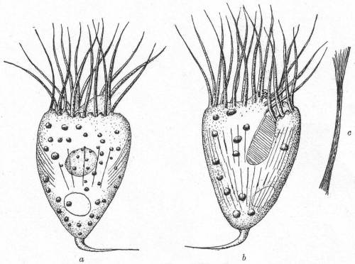
Although Fromentel's species is incompletely described, it is very evident that the organism corresponds fairly well with the Woods Hole variety. His was a fresh-water type; this is marine, but the caudal filament and the contractile vacuole are similar. Certainly in this case the organism can not be regarded as a Vorticella broken off its stalk, as Kent '81 suspected. The anterior process with its pigment spot; the cirri, the spherical nucleus, the position of the vacuole, etc., are all opposed to such an interpretation which Kent applied to the original species. Neither can it be a Tintinnoid. I place it provisionally as S. caudatum.
Key to the marine genera of Tintinnidæ.
Diagnostic characters: Body attached by a stalk to a cup. Inside the zone of membranelles is a ring of cilia (par-oral).
1. The test is gelatinous and more or less covered by foreign particles Genus Tintinnidium
2. The test is chitinous and clear. No foreign particles. Genus Tintinnus
3. The test is chitinous; covered by foreign particles, growth rings frequent Genus *Tintinnopsis
4. The test is chitinous, often covered by foreign particles. The test is marked by discoid, circular, or hexagonal spots. Genus Codonella
5. The test is perforated by pores of circular or hexagonal form. Genus Dictyocysta
* Presence at Woods Hole indicated by asterisk.
Genus TINTINNOPSIS Stein '67.
(Stein '67; Kent '81; Daday '87; Bütschli '88.)
Medium-sized ciliates, inclosed in a chitinous lorica with embedded sand crystals. The form of the house, or lorica, varies greatly. In some cases the mouth opening is wide, giving the lorica a bell form; it may be long and tubular, short and spherical, or variously indented. The animal is attached, as in the closely allied genus Tintinnus, by a peduncle to the bottom of the lorica. The anterior end of the animal is inclosed by two complete circles of cilia; one, the outer, forming the adoral zone, is composed of thick tentacle-like membranelles, the other consists of shorter cilia within the adoral zone. The mouth leads into a curved œsophagus containing rows of downward-directed cilia (Daday). The entire body is covered with cilia, but as the lorica is always opaque these can be made out only when the animal is induced to leave the house. The only difference between this genus and Tintinnus is the covering of foreign bodies—usually sand crystals. Movement is rapid and restless, and peculiarly vibratory, owing to the apparent awkwardness in moving the house. Salt water.
Tintinnopsis beroidea Stein, var. plagiostoma Daday. Fig. 47.
Fig. 47.— Tintinnopsis beroidea. ENLARGE
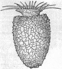
Synonym: Codonella beroidea Entz '84.
The shell is colorless, thimble-shaped, with a broadly rounded posterior end. The body is cylindrical. The internal organs were not observed. Membranelles 24 in number. Length 50µ; greatest diameter 40µ.
Var. compressa Daday '87.
The posterior end of the shell is pointed, the lower third of the shell is swollen, the upper third is uniform in diameter and without oral inflation or depression. Nucleus not seen.
Length 70µ; greatest diameter 48µ.
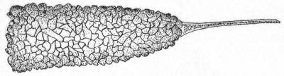
Tintinnopsis davidoffi Daday. Fig. 48.
The shell is large, elongated, and provided with a considerable spine. The chitin of the shell is covered with silicious particles of diverse size. The internal structures were not observed.
Length of shell and spine 230µ; diameter of the oral aperture 54µ.
Fig. 48.—Tintinnopsis davidoffi. ENLARGE
The variations of these species are considerable, and as the internal structures, such as the nucleus, are essential in fixing their systematic position, I place them as above, provisionally, and until further observations can be made.
Key to families of Hypotrichida.
a. Peristome indistinct; cilia on ventral surface uniform and not differentiated into cirri Family Peritromidæ One genus, *Peritromus
b. Peristome more or less indistinct; cilia reduced to a few rows on the ventral surface; anal and frontal cirri present Family Oxytrichidæ
c. Cilia entirely reduced; frontal and anal cirri present or reduced; macronucleus band-formed or spherical Family Euplotidæ
d. Peristome reduced to left edge and does not reach over the anterior margin Family Aspidiscidæ One genus, *Aspidisca
* Presence at Woods Hole indicated by asterisk.
Genus PERITROMUS Stein '62.
(Stein '62, '67; Maupas '83.)
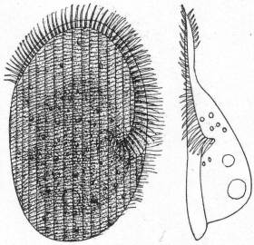
The body is flat, colorless or tinged with yellow, and contractile. It is elliptical in outline, with broadly rounded ends; in some cases the left edge is slightly incurved, the right edge convex. The ventral surface is flat, the dorsal surface is arched in the middle region of the body. The edges being flat are somewhat more transparent than the remainder of the body. The ventral surface is striated by longitudinal straight or slightly curved lines, the dorsal surface is smooth and without cilia. (Maupas describes bristles on the back, but this is not corroborated.) The adoral zone is fairly well developed, but not distinctly marked off from the remaining ventral surface. It begins on the right side and extends entirely around the frontal margin and down the left side below the middle of the body, where it turns suddenly to the right, entering the slightly insunk peristome. The mouth leads into a short, indistinct œsophagus. One contractile vacuole is situated in the dorsal swelling at the posterior end of the animal. Macronucleus double, one in each side of the dorsal swelling. Movement is slow and creeping, with a peculiar method of contracting the more hyaline edge, which may turn upward or around a foreign object.
Fresh (?) and salt water.
Peritromus emmæ Stein. Fig. 49.
With the characters of the genus.
Fig. 49.— Peritromus emmæ, ventral and lateral aspects. ENLARGE
Key to the marine genera of Oxytrichidæ.
Diagnostic characters: The peristome is not always marked off from the frontal area. In the most primitive forms the cilia on the ventral surface are similar to those of the preceding family (Peritromidæ). Usually some of the anterior and some of the posterior cilia are fused into cirri, distinguished as the frontal and anal cirri, respectively. In the majority of forms all of the cilia are thus differentiated; strong marginal cirri are formed in perfect rows, and ventral cirri in imperfect rows. In addition to the adoral zone there is an undulating membrane on the right side of the peristome, and in some cases a row of cilia between the membrane and the adoral zone. These are the par-oral cilia and they form the par-oral zone.
1. The posterior end is pointed or tail-like 2
The posterior end is rounded; not tail-like 5
2. The front end is pointed 3
The front end is rounded 4
3. Frontal and anal cirri absent; often tube-forming Genus Stichotricha
Eight frontal and 3 caudal cirri; not tubiculous Genus Gonostomum
4. Anal cirri present; with or without short lateral bristles Genus *Epiclintes
Anal cirri absent; no bristles Genus Uroleptus
5. With frontal cirri 6
No frontal cirri; 2 to 3 rows of ventral cirri; anal cirri small Genus Holosticha
6. Right margin of peristome straight as far as the anterior end; 5 rows ventral cirri; 5 anal cirri Genus Oxytricha
Right margin of peristome curved 7
7. Five rows or less of ventral cirri 8
More than 5 rows of ventral cirri Genus Urostyla
8. Membranelles normal; 5-10 anal cirri; no caudal cirri Genus *Amphisia
Membranelles normal; 5 to 10 anal cirri; 3 caudal cirri Genus Stylonychia
Membranelles very large and powerful; adoral zone not continued to mouth; 5 anal cirri Genus Actinotricha
* Presence at Woods Hole indicated by asterisk.
Genus EPICLINTES Stein '62.
(Stein '62, '64, '67; Mereschowsky '79; Gruber '87; Bütschli '88.)
Very active, contractile, colorless forms of rather small size. In the fully expanded condition the body is oval and long, with its greatest width in the center or at the front half of the body. The posterior end is always drawn out into a relatively long tail, which is extremely elastic. The peristome is short and stretches around the front end of the animal. In the frontal region are from one to three rows of cirri. The ventral surface is covered with longitudinal rows of cilia, the number of rows being in dispute (6 to 7 according to Stein; 9 according to Mereschowsky and Rees) Some of these cilia project from the lateral edges and from the posterior end, where they are slightly elongated. The anus is dorsal and placed at the beginning of the posterior process. Macronucleus probably double. Movement is rapid and restless, the tail process contracting to jerk the body backward. Salt water.
Fig. 50.— Epiclintes radiosa. ENLARGE
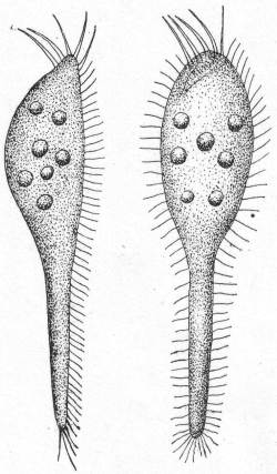
Epiclintes radiosa Quenn. Fig. 50.
Synonym: Metra radiosa Quenn.
The body is elongate, slightly narrowed anteriorly, and drawn out posteriorly into a long, retractile, tail-like portion. Five large cirri extend outward from the anterior extremity. The caudal portion may be extended to a distance equal to twice the length of the body or contracted to half the length. The peculiar nervousness of this form made it extremely difficult to study, and the oral region was imperfectly made out. The anterior cirri appear to line the upper left border of the peristome, which is marked by a row of large cilia. The peristome begins upon the right side of the anterior end and passes backward and to the left, narrowing at this point. The mouth is very small and difficult to see. It is apt to stay in one locality under zooglœa, switching back and forth with great vivacity, or hanging on by the posterior cilia while the anterior end stretches out in the surrounding medium. Nucleus and contractile vacuole were not observed. Length 45µ.
Genus AMPHISIA Sterki '78.
(Sterki '78; Kent '81; Bütschli '88.)
The body is plastic and soft, colorless or slightly tinged with yellow or red. In form it is oval or elongate, the posterior end is rounded and slightly reduced in diameter, but does not form a distinct tail. The anterior end is also rounded and similarly reduced in width. There are two rows of marginal cirri (Randcirren), which may be placed some distance from the edge, and two or three rows of ventral cirri between them. There are from 3 to 5 frontal cirri of larger size than those of the ventral rows, and from 5 to 10 anal cirri. (The genus Holosticha is similar in all respects save the presence of frontal cirri.) The macronucleus is double; the contractile vacuole is central and on the left side. The peristome is long and rather narrow and carries an undulating membrane on its right margin. Fresh and salt water.
Amphisia kessleri Wrzes. '77. Fig. 51.
Synonyms: Trichoda gibba Müller; Oxytricha gibba Stein '59; O. velox? Quen. '69; O. kessleri Wrzes. '77.
Body elongate, slightly sigmoid and swollen in the center, about 3-1/2 times as long as broad; the rounded anterior end is turned to the left, the similarly rounded posterior end to the right; both ends taper slightly. The peristome is long and narrow, with a distinct adoral zone which appears broken in its course. To the right of this adoral zone is a single line of preoral cilia. On the right border of the peristome is an undulating membrane. The three frontal cirri form a triangle and the five smaller anal cirri form a continuous line with the broken row of ventral cirri. There are two and one-half rows of ventral cirri and the marginal cirri are drawn in until they are ventral in position.
Length 135µ; greatest width 40µ.
Fig. 51.—Amphisia kessleri. ENLARGE
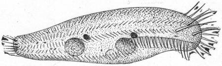
This variety differs from O. kessleri as described by Wrzesniowski in having three frontal cirri instead of four. Another difference is in the structure of the nuclei and in their position. These differences are too minute to warrant a specific name. O. velox of Quennerstedt is probably the same as 0. kessleri, but differs in having three complete rows of ventral cirri. O. velox has three frontal cirri in a line, thus differing from the Woods Hole form.
Key to the marine genera of Euplotidæ.
Diagnostic characters: Cilia, as well as the frontal, marginal, and ventral cirri, very much reduced; the anal cirri, on the other hand, are always present. The macronucleus is band-form.
1. Frontal cirri more than 8 2
Frontal cirri less than 8 3
2. Eleven marginal cirri on the left side; 11 frontal cirri Genus Certesia
Four marginal cirri, 2 on each side; 9 to 10 frontal cirri Genus *Euplotes
3. Seven frontal, 5 anal, 3 right marginal, and 2 left marginal cirri Genus *Diophrys
No frontal, 5 anal, 3 right, and 2 left marginal cirri Genus *Uronychia
* Presence at Woods Hole indicated by asterisk.
Genus EUPLOTES (Ehr. 1831) Stein '59.
(Ehrenberg '31, '38; Stein '59; Cl. & Lach. '58; Quennerstedt '65, '67, '69; Bütschli '88; Kent '81; Gourret & Roeser '88; Möbius '88.)
Small to medium-sized forms. Rigid in form, colorless, or green by chlorophyl. They are quite flat on the ventral surface but decidedly arched dorsally, and the contour is usually oval. The anterior end is broadly rounded to truncate; the posterior end is similarly rounded, or may be somewhat pointed. The mouth is placed centrally or near the left margin, and from it the right edge of the peristome forms a curved line to the left, which bends forward, thus making the greater part of the left edge the peristomial area. In front the peristome bends sharply to the right and extends as far as the right end of the adoral zone. Upon the frontal and median ventral surface are 9 to 10 great cirri (Bauchwimpern of Stein). Posteriorly five great anal cirri stretch out beyond the posterior body margin. In addition to these there are two smaller marginal cirri upon the left body edge, and two similar ones on the hinder part of the body. The dorsal surface is rarely smooth, but usually is marked by longitudinal ridges, and rows of dorsal bristles have been described. The single contractile vacuole lies on the right side in the region of the anal cirri, sometimes just above them, sometimes below. The anus is posterior and on the right side. The characteristic macronucleus is long and band-form, its main portion being usually on the left side with an anterior and a posterior arm toward the right. Movement is rapid swimming, which, however, is frequently broken by creeping periods, during which the animals appear to be examining the foreign body on which they creep.
Fresh and salt water.
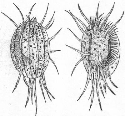
Euplotes charon Ehr. Fig. 52.
Synonyms: Trichoda charon Müller; Plœsconia charon; P. affinis, subrotunda, radiosa, longiremis, Dujardin '41.
The body is oval, small, and somewhat variable in length. The carapace is strongly marked upon the dorsal side by deep longitudinal grooves, 6 to 8 in number; the grooves may be absent, however. The adoral zone extends to the posterior third of the body, the mouth and œsophagus are directed anteriorly. There are 10 ventral cirri, 7 of which are on or near the frontal border and 3 near the right edge. There are 5 posterior cirri and 4 anal cirri, of much smaller size. The cirri may or may not be fimbriated, the latter condition indicating the approaching disintegration of the body and is abnormal. The macronucleus is long and band-formed or horseshoe shape. The contractile vacuole lies on the right side dorsal to the posterior cirri.
Fresh and salt water. Length 45µ; diameter 25µ.
Fig. 52.— Euplotes charon, dorsal and ventral aspects. ENLARGE
Euplotes harpa Stein. Fig. 53.
Fig. 53.— Euplotes harpa. ENLARGE
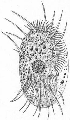
The body is elongate, oval, somewhat widened anteriorly, and has rounded ends. The frontal margin is three-toothed. Ten ventral cirri. Dorsal surface provided with 8 longitudinal markings. The peristome is long and broad, with considerable variation. The adoral zone consists of powerful membranelles arranged in a continuous curve from the mouth to the extreme right frontal margin. Seven of the 10 ventral cirri are situated at the anterior extremity; the remainder are arranged in a triangle on the right edge. The anal cirri, 5 in number, are long and stiff; the marginal cirri smaller and finer. The nucleus and contractile vacuole are similar to those of the preceding species.
Length 95µ; width 54µ.
Genus DIOPHRYS Dujardin '41.
(Bütschli '88.)
Medium size, colorless to yellow, rigid in form. The body contour is oval, the anterior end being rounded or slightly reduced, the posterior end usually cut in on the right side. The peristome is broad but less extensive than in Euplotes, and may extend beyond the middle of the body. Its right edge is convex toward the right side, extends forward and does not turn again to the right. The anterior ventral surface has 7 to 8 scattered cirri and just behind the mouth is a transverse row of large anal cirri. In the sharp in-cut of the posterior end are three great angular cirri. Two lateral cirri are placed on the left of the median line between the mouth and the anal cirri, and usually in a slight hollow. The contractile vacuole is on the right side in the vicinity of the anal cirri. The macronucleus is in two parts, each band-form, one anterior, the other posterior in position. Movement is rapid and steady. Salt water.
Diophrys (Styloplotes) appendiculatus Stein '59. Fig. 54.
Synonyms: Styloplotes appendiculatus Stein '59; Kent '81; Quennerstedt '67, etc.
Fig. 54.— Diophrys appendiculatus. ENLARGE
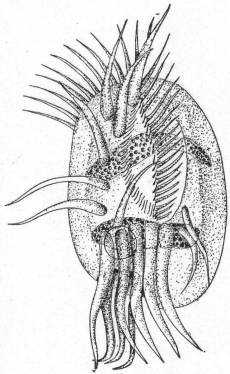
The general form resembles Euplotes. Its outline is oval and regular except at the posterior end on the right side, where there is a considerable indentation. The frontal margin is characterized by a row of powerful membranelles, which become smaller at the peristome and at the mouth they are of characteristically small size. The ventral cirri are 7 in number. Five of them are in one row from the anterior end down the right side nearly to the anal cirri; 1 is on the frontal border between the first two; 1 lies just anterior to the second anal cirrus from the right side. The 5 anal cirri are large and powerful and extend some distance beyond the posterior end of the body. In all specimens observed these cirri curve to the left. Dorsal to the anal cirri and placed deep into the dorsal pit are 3 large, sharply curved cirri, which in most cases are fimbriated, but when the specimens are normal these are pointed and curve abruptly to the right. Two smaller cirri lie to the left of the group of anal cirri. The peristome is well-marked by the adoral zone, and upon its right border there is a row of cilia, and a similar row of cilia runs along the base of the oral membranelle. The macronucleus is double and consists of two elongate cylindrical masses lying parallel with one another. One of these is in the anterior region; the other is posterior. The contractile vacuole lies dorsal to the anal cirri and anterior to the three dorsal cirri. The movement and general activities resemble those of Euplotes.
Length 50µ; diameter 25µ.
Genus URONYCHIA Stein '52.
(Stein '59, '67; Quennerstedt '67; Kent '81; Bütschli '88.)
Medium-sized colorless ciliates of usually constant body form. The body is somewhat short and oval in outline. The anterior end is broadly truncate, the posterior end rounded or slightly pointed. The ventral and dorsal surfaces are considerably arched and the latter usually has a number of rows of longitudinal stripes. The open peristome is broad and reaches back to the middle of the ventral surface and beyond. According to Stein, the two edges can approach each other, thus opening and closing the peristomial area. Its right edge forms a greater angle with the front edge than in the genus Euplotes, and the left edge forms a greater angle with the front edge than in that genus. The left edge also appears to cover over the adoral zone slightly. There are no ventral cirri in front, but on the posterior ventral surface are 7 great springing cirri. Five of these are inserted on the right aide in a deep in-sinking, and the other 2 in a similar depression on the left ventral surface. Above the 5 right-side cirri, i.e., dorsal to them, but in the same depression, are 3 angular cirri. A few edge cirri are found to the left of them and another to the right of the 5 cirri. The contractile vacuole is on the left side between the main groups of cirri. The macronucleus is band-form or spherical, and is situated in the middle region of the body. Movement consists in forward swimming with sudden springs.
Salt water.
Uronychia setigera, n. sp. Fig. 55.
This species is very common in the Woods Hole waters. It is small, colorless, and very active. The most characteristic feature is the posterior end with its relatively enormous cirri, which are apparently large enough for an animal four times its size. The form is ovoid, widened posteriorly.
Fig. 55.— Uronychia setigera. ENLARGE
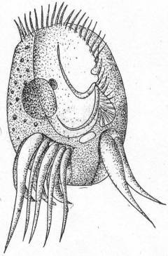
The ventral surface is flat and has two excavations in the posterior end. The right hollow is larger and contains 5 great cirri of unequal size, the extreme right one being the largest. The left hollow contains 2 cirri, also of dissimilar size. Dorsal to the 5 right cirri are 3 sickle-formed cirri, which are usually fimbriated. These are pointed and curve regularly to the left. The peristome is wide and open, and a small pocket-like hollow on its left border indicates the region of the mouth. The adoral zone runs into this pocket and the mouth is located in its lower right-hand corner. In U. transfuga the right border is generally described as having a membrane of extreme delicacy. I was unable to see such a membrane in this form, but in its place there are 2 flagella-like cirri extending from the margin of the mouth-opening into the peristome, and these vibrate slowly. I do not believe these could be the moving edge of an undulating membrane, for they are quite distinct. The macronucleus is spherical instead of band-form, and a single micronucleus is closely attached. This is unlike the European species U. transfuga, in which the nucleus is elongate. The contractile vacuole lies between the two sets of posterior cirri. There are no marginal folds like those of the European species.
Length 40µ; width 25µ. Common.
Genus ASPIDISCA Ehr. 1830.
(Perty '52; Cl & Lach. '58; Stein '59; Quennerstedt '65, '67, '69; Mereschowsky '79; Kent '81; Bütschli '88.)
Small, colorless, and rigid forms, with nearly circular to oval contour. The left side is usually straight, or at least but slightly convex. The right side is much more convex, and the right margin is considerably thickened. The ventral side is flat, the dorsal surface convex, with from one to several longitudinal ridges which run more or less parallel with the right edge. The peristome is limited to the left edge, where it forms a small depression which may or may not reach the anterior border, but which in no case runs around the anterior margin. The left peristome margin in some cases grows over the peristome depression toward the right, thus making a sort of cover for the peristome. In the posterior region is a deep depression, from which 5 to 12 cirri take their origin. Seven or 8 cirri are placed in the anterior half of the ventral surface and are arranged more or less in rows. The anus is on the right side in the region of the anal cirri (Stein). The contractile vacuole is generally on the right side and similarly located. The macronucleus is a horseshoe-shaped body. Movement rapid, somewhat in circles, and rather uniform.
Fresh and salt water.
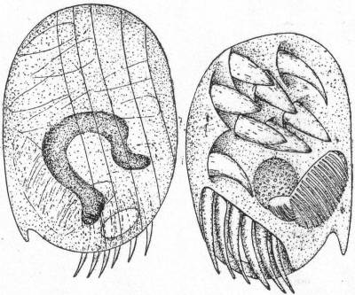
Aspidisca hexeris Quennerstedt '67. Fig. 56.
The carapace is elliptical, about 1-1/2 times as long as broad, rounded at the extremities. The left border of the carapace bears a spur-like projection. The ventral cirri are short and thick, and are very characteristic of the species. When moving slowly they look much like nicely-pointed paint brushes, but when the animal is compressed they quickly become fibrillated, and then look like extremely old and worn brushes. These cirri are placed in depressions in the ventral surface and each one appears to come from a specific shoulder. At the posterior end an oblique hollow bears 6 unequal cirri placed side by side. The extreme right cirrus is the largest, and they become progressively smaller to the opposite end. Dorsal to these lies the contractile vacuole. The peristome is in the posterior half of the body and an undulating membrane extends from it into the œsophagus. The dorsal surface is longitudinally striated by 5 or 6 lines, which are usually curved. The nucleus is horseshoe-shaped and lies in the posterior half of the body. Length 68µ; diameter 48µ.
Fig. 56.— Aspidisca hexeris. ENLARGE
This form was incorrectly mentioned as Mesodinium sp. by Peck '95:
In the figure given by Quennerstedt there are only 7 ventral cirri. In the Woods Hole form there are 8, 7 of which are anterior, 6 of them about one central one. The eighth cirrus is by itself, near the base of the largest posterior cirrus. These cirri, in spite of their size, are easily overlooked and more easily confused, but by using methylene blue they can be seen and counted.
Aspidisca polystyla Stein. Fig. 57.
Fig. 57.— Aspidisca polystyla. ENLARGE
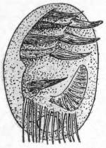
This species is similar to A. hexeris, but is smaller, very transparent, and without the spur-like process on the left edge of the carapace. The chief difference, however, lies in the number of anal cirri. These are 10 in number and they are arranged obliquely as in the preceding species, with the largest one on the right and the smallest on the left. The ventral cirri are 8 in number, and are arranged in two rows, one of which, the right, has 4 cirri closely arranged, the other having 3 cirri close together and one at some distance, near the largest anal cirrus. The peristome, contractile vacuole, and nucleus are similar to the preceding. Length 36µ; width 22µ.
Stein assigns only 7 ventral cirri to this species, but he also describes 2 very fine bristle like cilia (p. 125) and pictures them in figs. 18, 19, 20, and 21 of his Taf. III in the same relative position as my eighth cirrus. I am positive that cilia do not occur on the ventral face of this form, and that the characteristic cirri are the sole locomotor organs.
Key to families of Peritrichida.
a. Peristome drawn out into funnel-like process; parasitic Family Spirochonidæ
b. Adoral zone and circlet of cilia at opposite end. Adoral zone left-wound. Parasitic. Family Lichnophoridæ (one genus, *Lichnophora)
c. Adoral zone a left-wound spiral. Attached or unattached forms. Family Vorticellidæ
* Presence at Woods Hole indicated by asterisk.
Genus LICHNOPHORA Claparède '67.
(Gruber '84; Fabre-Domergue '88; Bütschli '88; Wallengren '94; Stevens 1901.)
Small or medium-sized colorless animals, extremely elastic and flexible. The anterior part, bearing the adoral zone, is round or oval in ventral view, and has a flat ventral and a highly arched dorsal surface. The posterior end of the animal is reduced to a stalk-like structure which is broadened at the extremity to form a sucking disk. The surface of this disk and the surface of the peristome may be brought into the same plane by the characteristic bending of the stalk portion. A ciliated girdle is placed at the edge of the sucking disk. A well-developed adoral zone incloses the peristome; it begins at the mouth on the left side and includes nearly all of the peristome in its left-wound spiral, the extremity approaching closely the end near the mouth. The macronucleus is a long-beaded structure, or it may be in several parts connected by strands (Gruber). The contractile vacuole is on the left side in the region of the mouth. Salt water.
Lichnophora macfarlandi Stevens. Fig. 58.
The body is elongate; oral disk variable in form, attachment disk clearly defined and constant. The stalk is very contractile and elastic, constantly changing in shape. When detached from the host the animal moves with a very irregular and indefinite motion. When attached it moves freely over the surface on its pedal disk. The latter is bordered by four membranes composed of cilia. A distinct axial fiber extends from the pedal disc to the peristome and gives off a number of branches. This fiber is analogous to the myonemes in Vorticella. An indistinct longitudinal furrow can be made out occasionally. The nucleus is in 5 or 6 separate pieces, of which 1 is found in the pedal disk and 1 or 2 in the neck.
On the egg capsules of Crepidula plana; also reported upon annelids at Woods Hole.
Length 60µ from disk to extremity of the peristomial disk.
Fig. 58.— Lichnophora macfarlandi. ENLARGE
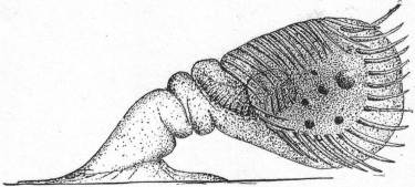
This form does not agree in all respects with Stevens's species, but the agreement is so close in other respects that I believe it can be safely identified as L. macfarlandi. The mode of life is different, and the macronucleus is different, there being from 25 to 30 fragments in Stevens's form and only 5 or 6 in the present one. There is, however, the same evidence of chain formation in both of them. The length of the oral cilia in Stevens's form is 18µ in fixed and 30µ in living forms. In the Woods Hole form the cilia are not more than half that length.
Key to the marine genera of Vorticellidæ.
Diagnostic characters: Attached or unattached forma of peritrichous ciliates in which the adoral zone seen from above forms a right-wound spiral. A secondary circlet of cilia around the posterior end may be present either permanently or periodically.
1. Posterior ciliated girdle permanent around an attaching disk 3
2. Posterior ciliated girdle, temporary during motile stage 4
3. Body cylindrical:
(a) With ring of stiff bristles above the ciliated girdle Genus Cyclochæta
(b) Without accessory ring of bristles; with velum Genus Trichodina
Body conical; general surface ciliated Genus Trichodinopsis
4. No test and no stalk Genus Scyphidia
5. No test; with stalk containing contractile thread 8
6. No test; with stalk but without contractile thread Genus Epistylis
7. With a test; with or without a stalk Genus *Cothurnia
8. Individuals solitary Genus *Vorticella
Individuals colonial; entire colony contractile Genus *Zoothamnium
Individuals colonial; parts only of the colony contractile Genus Carchesium
* Presence at Woods Hole indicated by asterisk.
Genus VORTICELLA (Linnæus 1767) Ehr. '38
(Bell Animalcule Leeuwenhoek 1675; Ehrenberg '38; Dujardin '41; Stein '51; Cl. & Lach. '58; Greeff '70; Bütschli '88; Kent '81; Stokes '88; etc.)
Medium-sized ciliates of general bell-like form. They may be colorless, or yellow and green through the presence of Zoochlorella. When not contracted, the peristome end is widespread, rarely narrowed. The adoral zone and peristome agree with the details given in the family characteristics. The chief character is the attachment of the posterior end by means of a single, longer or shorter, stalk, which contains a highly contractile thread easily distinguished in the living animal. Another character is the absence of colony formation. Contractile vacuole, single or double, usually connected with a sac-like reservoir. The macronucleus is invariably long and band-formed, with attached micronucleus. Fresh and salt water.
So many species of Vorticella have been described that the task of collecting data and of arranging the synonyms is extremely irksome and difficult. Stokes enumerates 66 species, inhabiting fresh and salt water, and several other new species have been added since his work. I am impressed with the fact that new species have been created without proper regard for the manifold variations which nearly all of the Ciliata show, and I believe the 66 species might be safely reduced to 12 or 15.
Vorticella patellina Müller. Fig. 59.
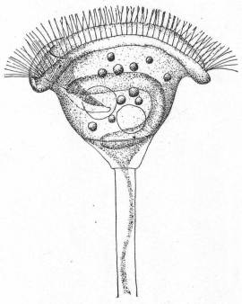
Body campanulate, widest at anterior border, from which it tapers directly to the pedicle. The diameter of the peristome is a little larger than the length of the body. The ciliary disk is but little elevated. The cuticle is not striated and the body plasm is quite transparent. Length 52µ.
Fig. 59.— Vorticella patellina. ENLARGE
Vorticella marina Greeff. Fig. 60.
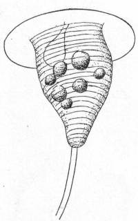
The body is conical but variable, and may he short or elongate, so that relative length and breadth offer no chance of identification. In general the body is campanulate. The distinguishing feature is the transverse annulation of the bell.
Small, but common, and grows in small social groups. Length 35µ.
Fig. 60.— Vorticella marina. ENLARGE
Genus ZOOTHAMNIUM (Bory de St. Vincent 1824) Stein '38, '54.
Fig. 61.— Zoothamnium elegans. ENLARGE
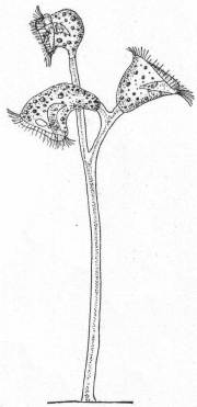
Colorless and highly contractile forms growing in small or large colonies. The form and structure of the individuals is not different from Vorticella. The colonies are usually richly branched upon the dichotomous plan and the entire colony is contractile. The main character is that with each division of the individual the stalk also divides, each daughter cell getting one-half of the parent stem. The stems therefore remain in communication, so that a simultaneous contraction results, and the colony as a whole is withdrawn. In some species so-called macrogonidia, or larger sexual individuals, are developed alongside the usual ones. Fresh and salt water.
Zoothamnium elegans D'Udekem '64? Fig. 61.
The bodies are variable—peristomial border widely dilated, tapering and attenuate posteriorly. The pedicle is slender, smooth, and transparent, and branches sparsely at its distal extremity. There are but few zooids (3 to 4). The ciliary disk projects conspicuously beyond the peristomial border. The pharyngeal cleft is very distinct and extends beyond center of body. Length of body 80µ.
Genus COTHURNIA (Ehr. '31) Clap. & Lach. '58.
Colorless forms of medium size-in some cases they may be green by Zoochlorella. The general structure is similar to that of Vorticella, but the individuals are elongate and occupy houses. The macronucleus is invariably long and band-form. The distinguishing character is the colorless or brownish lorica of quite variable form but always attached. These houses may be finger-formed, with widened center, or widened mouth, or constricted mouth, and the like. Ring-formed swellings are frequently developed. Sometimes the mouth becomes twisted and the lorica is therefore bilateral. The houses are attached either directly to some foreign object or by means of a short stalk. The animals are similarly fastened to the lorica, sometimes directly, sometimes by means of a short stalk. When they contract they draw back to the bottom of the lorica; when expanded they usually stretch out of the mouth opening. In some forms there is an operculum, by means of which the opening of the shell can be closed when the animal is retracted. Fresh and salt water.
The number of species of Cothurnia has become so great that the difficulty in placing forms is almost sufficient to discourage the systematist; as Bütschli well remarks, the variations in the theca have been made the basis of new species so many times that the genus is almost as confused as Difflugia among the rhizopods or Campanularia among the hydroids. The length of cup, of stalk, the presence of annulations on stalk or cup, etc., have given rise to many specific names, the majority of which I believe can be discarded. According to such differentials the same branch of an alga holding a hundred specimens of Cothurnia crystallina yield 10 or 12 species, whereas they are merely growth stages of one and the same form.
Cothurnia crystallina Ehr. Fig. 62.
Synonyms: Vaginicolla crystallina Ehr., Perty, Eichwald; V. grandis Perty; V. pedunculata Eichwald; Cothurnia crystallina Claparède & Lachmann, D'Udek.; C. gigantea D'Udek; C. maritima, C. crystallina Cohn; C. grandis Meresch.
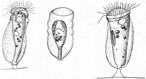
The form of the cup shows the greatest differences; sometimes it is cylindrical, sometimes elongate thimble-shape, sometimes pouch-shape, corrugated or smooth on the sides, and wavy or smooth on border. Frequently the basal part becomes stalk-like, but this is very short. When present, the stalk may or may not have a knob-like swelling. The animal within the cup may or may not be borne on a stalk, and this stalk may or may not be knobbed. The cups are colorless or brown. The animal is very contractile and may stretch half its length out of the cup or retract well into it. There is no operculum. The length of the cup varies from 70µ to 200µ (C. gigantea; Vag. grandis, etc.). From Entz.
There is nothing to add to Entz's characterization of this species, which is found both in fresh and salt water. The variability of the cup and stalk is quite noticeable in the Woods Hole forms.
Fig. 62.—Cothurnia crystallina. ENLARGE
Cothurnia imberbis Ehrenberg, var. curvula Entz. Fig. 63.
Synonyms: C. imberbis Kent et al.; C. curvula Entz; C. socialis Gruber?
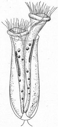
The lorica is swollen posteriorly, narrowest at the oral margin, bent on its axis and is supported on a short stalk. It is perfectly smooth and without annulations. The animal itself has no definite stalk. When fully expanded the animal emerges but slightly from the margin of the cup. Fresh and salt water. On red algæ. Dimensions of Woods Hole form: Cup 50 to 55µ long; greatest diameter 22µ; length of stalk 4 to 5µ.
Fig. 63.—Cothurnia imberbis. ENLARGE
Cothurnia nodosa Claparède & Lachmann. Fig. 64.
A. Smooth cup.—Cothurnia maritima Ehr., Eichwald, Stein, Kent.
B. Cross-ringed cup.—C. pupa Eichwald, Stein, Cohn; C. nodosa Cl & L.; V. crystallina Entz '78; C. pontica Meresch., Kent; C. cohnii and pupa Kent; C. longipes Kellicott '94.
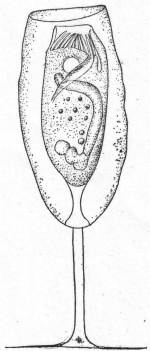
The cup is elongated, swollen centrally, tapering at oral end and conical at base or rounded. Oral opening either circular or elliptical. Cross rings may or may not be present, and the cup is either smooth or annulate. Length of cup 70µ to 80µ. The stalk which supports the cup is extremely variable in length. The animal is borne upon a stalk of variable length within the cup.
Entz states that the many variations which this species exhibits run into each other so gradually that he does not believe it wise to separate them. The Woods Hole forms which I found on algæ of various kinds were nearly of a size, and did not vary much from the one figured. Kellicott '94 described a Cothurnia from Woods Hole under the name of C. longipes, which I believe is only a long-stemmed variety of C. nodosa. My form has the following dimensions: Cup 75µ; cup stalk 38µ; animal stalk 14µ.
Fig. 64.—Cothurnia nodosa. ENLARGE
Key to families of Suctoria.
a. Unattached forms; ventral cilia present; one suctorial tentacle Hypocomidæ
b. Attached forms; thecate and athecate tentacles simple, one or two in number Urnulidæ
c. Thecate; posterior end of cup drawn out into stalk; walls perforated for exit of tentacles Metacinetidæ
d. Stalked or unstalked; globular; tentacles of different kinds, some knobbed, others pointed Podophryidæ (2 genera *Ephelota, *Podophrya)
e. Naked or thecate; stalked or not; tentacles numerous, usually knobbed and all alike Acinetidæ
f. Naked; athecate; tentacles numerous, all alike, knobbed and grouped in tufts. They may be simple or branched. Dendrosomidæ
g. Sessile forms resting on basal surface or on a portion raised like a stalk; tentacles many; short and knobbed; distributed on apical surface or localized on branched arms Dendrocometidæ
h. Stalked or sessile; tentacles long, rarely knobbed, supported on proboscis-like processes Ophryodendridæ
* Presence at Woods Hole indicated by asterisk.
Genus PODOPHRYA Ehr. '33.
(Bütschli '88; Stein '59; Perty '52; Cienkowsky '55; Quenn. '69; Hertwig '77; Maupas '81.)
Fig. 65.— Podophrya gracilis. ENLARGE
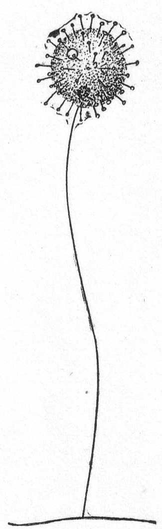
The body is globular, with tentacles radiating in all directions. The tentacles may be very short or very long. The stalk also is either short or long, and some species form stalks but rarely (P. libera). The macronucleus is centrally placed and globular to ovoid in form. The contractile vacuole is usually single. Reproduction takes place by division; the distal half developing cilia and becoming a swarm-spore. Fresh and salt water.
Podophrya gracilis, n. sp. Fig. 65.
Of all the Podophrya that have been described not one approaches this minute form in the relative length of the stalk. The body is spherical and is covered with short capitate tentacles. The stalk is extremely slender, bent, and without obvious structure. There are one or two contractile vacuoles in the distal half of the body. The nucleus is small and is situated near the insertion-point of the stalk. Reproduction not observed. Diameter of body 8µ; length of stalk 40µ. Only one specimen seen.
Genus EPHELOTA Str. Wright '78.
(Bütschli '88; Ishikawa '96; Sand '98.)
Small to medium-sized and large forms; colorless to brown. The body is globular or oval or wedge-shape, sometimes quadrangular. The stalk is variable, sometimes 1 mm. in length. The diameter of the stalk increases from the point of attachment to the body of the animal; it is usually striated either longitudinally or transversely, or both. The tentacles are of two kinds and are usually confined to the anterior half of the body. Some are long and sharp-pointed and adapted for piercing; others are short, cylindrical, usually retracted and capitate, adapted for sucking. Contractile vacuoles vary from one to many. The macronucleus is nearly central in position and usually of horseshoe shape, but is frequently branched and irregular. Reproduction is accomplished by external multiple budding, usually from the anterior half of the body. Salt water.
Ephelota coronata Str. Wright. Fig. 66.
Synonyms: Hemiophrya gemmipara S. K.; Podophrya gemmipara Hertwig.
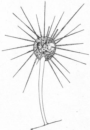
The body is spheroidal, ovate, or pyriform, with numerous sharp-pointed tentacles and a few straight, uniform tentacles. The stalk is about three times the length of the body and tapers from its widest part at the insertion in the body to the narrowest part at the point of attachment. It may or may not be longitudinally striated. This is one of the commonest of the Suctoria found at Woods Hole. It is usually present on Campanularian hydroids, but may be found on algæ and Bryozoa.
Length of body 90µ to 200µ.
Fig. 66.— Ephelota coronata. ENLARGE
Genus ACINETA Ehr. '33, Bütschli '88.
(Stein '54, '59; Claparède & Lachman '58; Quennerstedt '67; Hertwig '76; Mereschowsky '79; Entz '84; Kent '81; Maupas '83; Gruber '84; Gourret & Roeser '86, and others.)
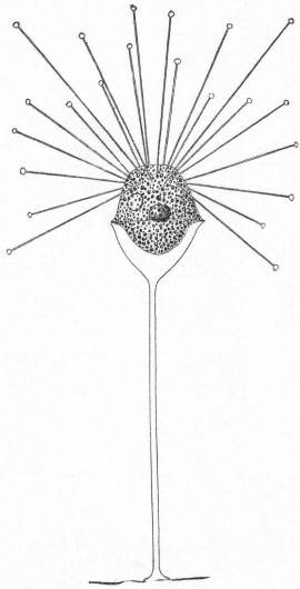
Fig. 67.—Acineta divisa. ENLARGE
Small to medium-sized forms. The distinguishing feature is that the stalk is swollen at the distal extremity to form a cup or basin in which the animal rests. The cup may be developed until the body is nearly inclosed. The macronucleus is spherical or band form. The contractile vacuole is usually single. Budding, so far as known, is endogenous. Fresh and salt water.
Acineta divisa Fraipont '79. Fig. 67.
This extremely graceful form is common on Bryozoa at Woods Hole. The cup is shaped like a wine glass and is specifically characterized by a cup-formed membrane upon which the animal rests. The animal thus has the appearance of being suspended on the edge of the cup. The stalk is slender and about 4 times the length of the body. The tentacles are all capitate and distributed, and about 2-1/2 times the body length. They sway back and forth very slowly. The nucleus is spherical and central in position. The contractile vacuole lies near the periphery.
Length of body 27µ; of stalk 100µ; of extended tentacle 65µ.
Acineta tuberosa Ehr. Fig. 68.
Fig. 68.— Acineta tuberosa. ENLARGE
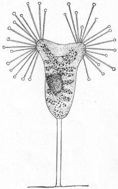
Large forms of Suctoria with tentacles arranged in fascicles. The stalk is variable in length and the cup is frequently so delicate that it can barely be made out. A specific characteristic is the break in continuity of the cup at different points, and through these places the tentacles emerge in bundles. The tentacles are capitate and in the Woods Hole form, 15 in number in each of the two bundles. The endoplasm is granular and yellowish in color. The coloring matter is frequently arranged in patterns. The nucleus is spheroidal. The contractile vacuole is in the anterior third of the body about midway between the bundles of tentacles. Reproduction not observed.
Length of body 330µ.
Genus TRICHOPHRYA Clap. & Lach. '58.
(See Kent '81; Entz '84; Bütschli '88; Sand 1901.)
Small forms to medium size; no cups or stalks. The body is spherical to elongate, usually, however, more or less irregularly lobed and changeable. The tentacles are in fascicles which are usually borne upon lobed or swollen places. The body is always more or less spread out. Contractile vacuoles variable. The macronucleus is spherical, elongate, band-formed or horseshoe-shaped. Reproduction takes place by endogenous budding, and the swarm spores are flat or lenticular with a distinct ciliary girdle. They are frequently parasitic. Fresh and salt water.
Trichophrya salparum Entz '84. Fig. 69.
Bütschli '88; Schewiakoff '93; Trichophrya ascidiarum Lachmann '59; René Sand 1901.
The body is somewhat cup-form, with a large, flat base. The anterior border is rounded, each of the ends being somewhat truncate and carrying a bundle of tentacles all capitate and similar. These may be continued internally as far as the nucleus (Sand). The cytoplasm is uncolored, but may contain some brilliant granules. The nucleus is granular, and spherical, band or horseshoe formed.
Fig. 69.— Trichophrya salparum. ENLARGE
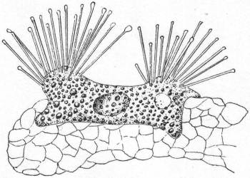
This species was found by Dr. G. Hunter on the branchial bars of the Ascidian Molgula manhattensis, where great numbers of them are often parasitic.
LIST OF REFERENCES.
AUERBACH, L. '54. Ueber Encystierung von Oxytricha pellionella. Zeit. wiss. Zool., V, 1854.
–––– '55. Ueber die Einzelligkeit der Amoeben. Zeit. wiss. Zool., VII, 1855.
BALBIANI E. G. '61. Recherches sur les phénomènes sexuelles des Infusoires. Jour. de la physiol:, IV, 1861.
–––– '85. Sur un Infusoire parasite du sang de l'Aselle aquatique. Rec. zool. Suisse, II, 1885.
BERGH, R. S. '79. Tiarina fusus Cl. & Lach. Vid. Med. f. d. Nat. Foren. Kjobenhavn.
–––– '82. Der Organismus der Cilioflagellaten. Morph. Jahr., VII, p. 177.
BRADY, H. B. '79. Report on the Foraminifera dredged by H. M. S. Challenger during the years 1873-1876. Challenger Reports, Zoology, IX.
BRANDT, K. '96. Die Tintinnodeen. Bibliot. Zoolog., XX, 1896.
BÜTSCHLI, O. '76. Studien über die ersten Entwickelungsvorg. der Eizelle, die Zelltheilung, und die Conjugation der Infusorien. Abhand. der Senck. naturf. Ges. Freiburg, X, 1876.
–––– '83-'88. Die Protozoen. Bronn's Klassen u. Ord. des Tierreichs.
CARPENTER, W. B. '56. Researches on the Foraminifera. Phil. Trans. (2), p. 547.
CARTER, H. J. '56. Notes on the fresh-water Infusoria of the island of Bombay. Ann. Mag. Nat. Hist. (2), XVIII.
–––– '63. On the coloring matter of the Red Sea. Id. (3), XI.
–––– '63a. On Amœba princeps and its reproductive cells. Id. (3), XII.
–––– '64. On fresh-water Rhizopods of England and India. Id. (3), XIII, p. 13.
–––– '65. On the fresh- and salt-water Rhizopods of England and India. Id. (3), XV, p. 277.
CIENKOWSKY, L. '55. Bemerkungen über Stein's Acinetenlehre. Bull. Phys. Math. Acad., St. Petersburg, XIII, p. 297 (also in Ann. Mag. Nat. Hist. (3), XIII).
–––– '55. Ueber Cystenbildung bei Infusorien. Zeit. wiss. Zool., VI, p. 301.
–––– '61. Beiträge zur Kenntniss der Monaden. Arch. f. mik. Anat., I, 1865, p. 203.
CLAPARÈDE, E. '54. On Actinophrys sol. Ann. Mag. Nat. Hist. (2), XV, 1854.
–––– '60. Recherches sur les annélides, etc., observés dans les Hébrides. Mém. Soc. phys. d'hist. nat., Genève, XVI, 1860.
–––– '67. Miscellanées zoologiques. Ann. d. sc. nat. zool. (5), VIII, 1867.
CLAPARÈDE ET LACHMANN '58-'60. Etudes sur les Infusoires et les Rhizopodes. Mém. Inst. genèvoise, V, VI, VII.
COHN, F. '54. Beiträge z. Entwicklungsges. der Infusorien. Zeit. wiss. Zool. III, 1851; IV, 1853.
–––– '57. Ueber Fortpflanzung von Nassula elegans Ehr. Zeit. wiss. Zool., IX, 1857, p. 143.
COSTE, M. '64. Développement des Infusoires ciliés dans une macération de foin. Ann. d. ac. nat. zool.(5), II.
DADAY, E. v. '86. Ein kleiner Beitrag der Infusorien-Fauna des Golfes v. Neapel. Mitt. Zool. St. Neap., VI, p. 481.
–––– '88. Monographie der Familie der Tintinnodeen. Mitt. Zool. St. Neap., VIII, p. 473.
DAVIS, J. '79. On a new species of Cothurnia. Jour. Roy. Mic. Soc., II, 1879.
DUJARDIN, F. '35. Recherches sur les organismes inférieures. Ann. d. sc. nat. zool. (2), IV, p. 343.
–––– '41. Histoire naturelle des Zoophytes infusoires. Paris, 1841.
EHRENBERG, C. G. '31. Ueber die Entwickelung und Lebensdauer d. Infusionsthiere. Abhand. d. Berlin Ak., 1831, p. 1.
–––– '38. Die Infusionsthierchen als vollkommne Organismen. Leipzig, 1838.
ENGELMANN, Th. W. '78. Zur Physiologie der Contractilen Vacuolen der Infusionsthiere. Zool. Anz., I, p. 121.
ENTZ, G. '84. Ueber Infusorien d. Golfes v. Neapel. Mitt. Zool. St. Neap., V, p. 289.
–––– '85. Zur naheren Kenntniss der Tintinnodeen. Mitt. Zool. St. Neap., VI, p. 185.
FABRE-DOMERGUE, P. '85. Note sur les Infusoires ciliés de la baie de Concarneau. Jour. d. l'anat. et de la phys., XXI, p. 554.
–––– '91. Matériaux pour servir a l'histoire des Infusoires ciliés. Ann. d. mic., III, 1890-91, p. 49.
FOL, H. '83. Further contribution to the knowledge of the Tintinnodea. Ann. Mag. Nat. Hist. (5), XII, p.73.
FRANCÉ, R. H. '97. Der organismus der Craspedomonaden. Budapest.
FROMENTEL. E. DE. '74. Etudes sur les microzoaires, etc. Paris, 1874.
GOURRET ET ROESER. '88. Contributions à l'étude des Protozoaires de la Corse. Arch. d. biol., VIII p. 139.
GREEFF, R. '66. Ueber einige in der Erde lebende Amoeben und andere Rhizopoden. Arch. f. mik. Anat., II, p. 299.
–––– '70. Untersuchungen ueber den Bau u. d. Naturgeschichte d. Vorticellen. Arch. f. Naturges., XXXVI, XXXVII.
–––– '71. Ueber die Actinophryens oder Sonnenthierchen des Süssenwassers als echte Radiolarien. Sitz. Ber. d. Niederrh. Ges. i. Bonn, XXVIII, p. 4.
–––– '75. Ueber Radiolarien u. Radiolarienartige Rhizopoden des Süssenwassers 2. Arch. f. mik. Anat., XI.
–––– '88. Studien über Protozoen. Sitz. Ber. Ges. z. Bef. d. ges. Nat., Marburg, 1888, p. 91.
GRENACHER, H. '68. Ueber Actinophrys sol Ehr. Verh. phys.-med. Ges. Freiburg (n. F.), I.
GRUBER, A. '83. Beobachtungen an Chilodon curvidentis n. sp. Festschr. 56. Vers. Deutsch. Naturf. gewid. v. d. Naturf. Ges. Freiburg, II, p. 1.
–––– '84. Die Protozoen des Hafens v. Genua. Nova Act. d. K. Leop.-Car. Deutsch. Akad. d. Naturf., XLVI, p. 475.
–––– '87. Ueber der Bedeutung der Conjugation bei den Infusorien. Ber. d. Naturf. Ges. Freiburg, II, p. 31.
HÆCKEL, E. '73. Zur Morphologie der Infusorien. Jena Zeit., VII, p. 516.
HERTWIG, R. '76. Ueber Podophrya gemmipara, nebst Bemerkungen zum Bau u. d. systemat. Stellung d. Acineten. Morph. Jahr., IV, p. 20.
HERTWIG U. LESSER. Ueber Rhizopoden u. denselben nahe stehende Organismen. Arch. F. mik. Anat., X, Suppl., p. 35.
HUXLEY, T. H. '57. On Dysteria, a new genus of Infusoria. Jour. Mic. Sci., V, p. 78.
ISHIKAWA, C. Ueber eine in Misaki vorkommende Art v. Ephelota, etc. Journ. Coll. Sci. Imp. Univ. Tokyo, Japan, X, pt. 2.
JAMES-CLARK, H. '66. On the Spongiæ ciliatæ as Infusoria flagellata, etc. Mem. Bost. Soc. Nat. Hist. (3), I, p. 1.
KENT, W. SAVILLE. '81. Manual of the Infusoria. London, 1881.
KLEBS, G. '84. Ein kleiner Beitrag z. Kenntniss d. Peridineen. Bot. Zeit., XLII, p. 721.
–––– '92. Flagellaten Studien 1. Zeit. wiss. Zool., LV.
LABBÉ, A. '95. Sur les Protozoaires marins de Roscoff. Arch. d. zool. expér. (3), N. et R., p. XIV.
LAUTERBORN, R. '94. Beiträge z. Süsswasserfauna der Insel Helgoland. Wiss. Meeresunt. Komm. wiss. Unt. d. Meere Kiel (2), I, p. 215.
LEIDY, J. '77. Remarks upon Rhizopods and notice of a new form. Proc. Ac. Sci. Phila., 1877, p. 293.
–––– '79. Fresh water Rhizopods of North America. Washington, 1879.
LIEBERKÜHN, N. '56. Ueber Protozoen. Notes from a letter to C. Th. v. Siebold. Zeit. wiss. Zool., VIII, p. 307.
MAUPAS, E. '81. Contributions à l'étude des Acinétiens. Arch. d. zool. expér. (1), IX, p. 299.
–––– '83. Contributions à l'étude morphologique et anatomique des Infusoires ciliés. Id. (2), I.
–––– '83a. Les sucto-ciliés de M. Mereschowsky. Comp. Ren., XCV, p. 1381.
–––– '88. Recherches expérimentales sur la multiplication des Infusoires ciliés. Arch. d. zool. exper. (2), VI, p. 165.
MERESKOWSKY, C. '78. Studien über Protozoen des nordlichen Russland. Arch. f. mik. Anat., XVI, p. 163.
–––– '81. On some new or little-known Infusoria. Ann. Mag. Nat. Hist. (5), VII, 1881.
MOEBIUS, K. '88. Bruchstücke einer Infusorienfauna der Kieler Bucht. Arch. f. Naturg., 1888.
PECK, J. I. '93. On the food of the menhaden. Bull. U. S. Fish Com., 1893, p. 113.
–––– '95. The sources of marine food. Bull. U. S. Fish Com., 1895, p. 351.
PERTY, M. '49. Mikrosk. Organ. der Alpen u. d. Italien. Schweiz. Mitt. d. Naturf. Ges. in Bern, 164-165.
–––– '52. Zur Kenntniss kleinster Lebensformen, etc., Bern.
POUCHET, G. '83; '85. Contributions à l'histoire des Péridiniens marins. Jour. de l'anat. et de la phys., XIX, XXI.
QUENNERSTEDT, A. '65; '67; '69. Bidrag till Sveriges Infusorie-fauna. Lunds Univ. Ärsskrift, II, IV, VI.
SAND, R. 1901. Etude monographique sur le groupe des Infusoires tentaculifères. Ann. d. la Soc. belge de microscopie, XXIV, XXV, XXVI.
SCHAUDINN, F. '95. Die Heliozoen. Das Tierreich, 1895.
SCHEWIAKOFF, W. '89. Beiträge z. Kenntniss der Holotrichen Ciliaten. Bib. Zool., V, p. 1.
–––– '93. Ueber einige ecto- u. ento-parasitische Protozoen der Cyclopiden. Bull. Soc. nat. Moscou, 1893. I.
SCHUETT, F. '95. Die Peridineen d. Plankton-Expedition, 1. Kiel u. Leipzig.
SCHULTZE, F. E. '74; 75. Rhizopodenstudien. Arch. f. mik. Anat., X, XI, XIII.
SCHULTZE, M. '62. Ueber d. Organismus d. Polythalamien. Leipzig, 1862.
SHEVYAKOV ?. '96. Monograph on Holotrichous ciliates. (In Russian.) Mem. of the St. Petersburg Acad., VII.
STEIN, F. '59; '78; '83. Der Organismus der Infusionsthiere. I. Infusoria, '59; II. Infusoria, '78; III. Flagellata, '83.
–––– '49. Untersuchung über die Entwicklungs d. Infusorien. Arch. f. Natur., I, p. 92.
–––– '54. Die Infusionsthiere auf ihre Entwicklungsgeschichte untersucht. Leipzig, 1854.
–––– '60. Ueber Leucophrys patula u. über 2 neue Infusoriengattungen Gyrocoris u. Lophomonas. Sitz. Ber. d. K.-böhm. Ges. d. Wiss. d. Prag, 1860, p. 4.
–––– '64. Ueber die neue Gattung Epiclintes. Id., 1864, I.
STERKI, V. '78. Beiträge z. Morphologie der Oxytrichinen. Zeit. wiss. Zool., XXXI, p. 29.
–––– '98. On the classification of ciliate Infusoria. Amer. Natur., XXXII, p. 425.
STEVENS, N. M. 1901. Studies on ciliate Infusoria. Proc. Calif. Acad. Sciences, III, 1.
STOKES, A. C. '84. Notices of some new parasitic Infusoria. Amer. Nat., XVIII, p. 1081.
–––– '85. Some apparently undescribed Infusoria from fresh water. Id., XIX, p. 18.
–––– '87. Some new hypotrichous Infusoria from American fresh waters. Ann. Mag. Nat. Hist. (5), 20.
TATEM, T. G. '67. New species of microscopic animals. Q. J. M. S. (n.s.), VII, p. 251.
VEJDOWSKY, F. '79. Monographie der Enchytraeiden. Prag, 1869.
WALLENGREN, H. '94. Studier ofver ciliata Infusorier, 1. Slagtet Lichnophora. Lund, 1894.
WALLICH, G. C. '63. Observations on an undescribed indigenous Amœba. Ann. Mag. Nat. Hist. (3), XI, XII.
WESTON, J. '56. On the Actinophrys sol. Q. J. M. S., IV, p. 116.
WRZESNIOWSKI A. '61. Observations sur quelques Infusoires. Ann. d. sc. nat. zool. (6), XVI.
–––– '69. Ein Beitrag zur Anatomie der Infusorien. Arch. f. mik. Anat., V, p. 25.
–––– '70. Beobachtungen über Infusorien a. d. Umgebung v. Warschau. Zeit. wiss. Zool., XX, p. 467.