TABLES.
Norman Walker translation
TABLE I.
SHOWING THE FREQUENT COMPLICATION OF NODULAR LEPROSY WITH TUBERCULOSIS.
No. Duration of Disease in Years. Lungs and Pleura. Liver. Spleen. Kidneys. Intestinal Canal.
1 11 Normal. Amyloid and leprous. Amyloid and leprous. Fatty degeneration. Amyloid.
2 6 Left-sided tubercular pleurisy; in lower lobes, lobular pneumonia. Leprous. Tubercular. Fatty degeneration. Normal.
3 10 Hydro-thorax. Amyloid and leprous. Leprous. ? Normal.
4 5 Cavities in both apices; tubercular peri-bronchitis in the upper lobes. Leprous. Amyloid. Amyloid. Amyloid and tubercular ulceration.
5 12 Normal. Amyloid. Amyloid. Amyloid. Normal.
6 8 Lobular pneumonia in the right lung. Leprous. Leprous. Fatty degeneration. Normal.
7 13 Lungs œdematous. Leprous. Leprous. Fatty degeneration. Normal.
8 7 In both lower lobes pneumonia, with commencing cavity formation. Leprous. Leprous. Normal. Normal.
9 5 In the left lung, cavities in the apex, and widespread lobular pneumonia. The same without cavity formation in the R. Amyloid and leprous. Leprous. Amyloid and fatty degeneration. Amyloid.
10 6 Œdema. Leprous. Leprous. Fatty degeneration. Normal.
11 5 In upper lobes, cavities, and lobular pneumonia. Amyloid and leprous. Amyloid and leprous. Amyloid. Amyloid.
12 11 Normal. Leprous. Leprous. Interstitial nephritis. Normal.
13 9 Cavities, tubercular peri-bronchitis, pleural tuberculosis. Amyloid & leprous (?). Leprous (?). Amyloid and tubercular. Amyloid and tubercular, tubercular peritonitis.
14 10 Normal. Amyloid and leprous. Leprous. Amyloid. Amyloid ulceration in the ileum.
15 14 Tubercular peri-bronchitis and pleurisy. Tubercular. Tubercular. Amyloid and tubercular. Amyloid and tuberculosis of omentum and mesentery.
16 4 Tubercular peri-bronchitis and pleurisy. Tubercular, amyloid, and leprous. Tubercular and amyloid and leprous. Tubercular and amyloid. Amyloid, tubercular ulceration, mesenteric tuberculosis.
17 7 Normal. Leprous and amyloid. Leprous and amyloid. Amyloid. Amyloid.
18 10 Numerous cavities and gelatinous pneumonia. Leprous and amyloid. Leprous and amyloid. Fatty degeneration. Peyer’s patches and solitary glands tubercular with commencing ulceration.
19 6 Numerous cavities and gelatinous pneumonia. Leprous. Leprous. Interstitial nephritis. Ulcerating plaques and solitary glands.
20 15 Normal. Fatty. Amyloid. Amyloid. Amyloid.
21 2 Normal. Leprous. Leprous. Normal. Normal.
22 14 A small pneumonia in R. Leprous. Leprous. Amyloid and fatty degeneration. Amyloid.
23 7 Cavities, tubercular peri-bronchitis, and lobular pneumonia in left lung. Leprous. Leprous and amyloid. Amyloid and fatty degeneration. Tubercular ulceration.
24 13 Bronchiectasis and lobular pneumonia. Leprous. Leprous. Amyloid and fatty degeneration. Normal.
25 8 Bronchiectasis and lobular pneumonia. Leprous. Leprous. Slight fatty degeneration. Normal.
26 5 Normal. Leprous. Leprous. Slight fatty degeneration. Normal.
27 ? Small cavities and lobular pneumonia. Leprous and amyloid. Leprous and amyloid. Amyloid. Normal.
28 3 Small cavities and lobular pneumonia. Leprous and tubercular. Leprous and tubercular. Fatty degeneration and amyloid. Amyloid ulcerated follicles and plaques.
29 1½ Tuberculosis of lungs and pleura. Leprous and tubercular. Leprous and tubercular. Acute nephritis, tuberculosis in R. kidney. Normal.
30 4 Caseous areas in both lungs. Leprous. Leprous. R. tubercular, L. hydronephrosis, tubercular cystitis. Normal.
31 6 Œdema, bronchitis. Amyloid. Amyloid. Amyloid. Amyloid.
32 24 Lobular pneumonia. Amyloid. Amyloid. Amyloid and fatty degeneration. Amyloid.
33 6 Cavities and lobular pneumonia. Leprous and amyloid. Leprous and amyloid. Normal. Amyloid.
34 5 Normal. Leprous. Leprous and amyloid. Amyloid and interstitial nephritis. Amyloid.
35 4 Pleuritic adhesions. Leprous. Leprous. Normal. Peritonitis, ulceration of the vermiform process.
36 14 Œdema. Amyloid. Normal. Amyloid and fatty degeneration. Amyloid.
37 4 L. Tubercular pleurisy; R. tubercular peri-bronchitis. Amyloid. Amyloid and tubercular. Tubercular. Amyloid, tubercular peritonitis.
38 6 Tubercular peri-bronchitis and lobular pneumonia; tubercular pleurisy in left lung. Leprous. Leprous. Normal. Amyloid, localised tubercular peritonitis around the spleen.
39 6 Tubercular peri-bronchitis, lobular pneumonia and small cavities. Leprous and amyloid. Leprous and amyloid. Tubercular and amyloid. Amyloid.
40 7 Normal. Leprous and amyloid. Leprous and amyloid. Amyloid. Normal.
41 8 Lobular pneumonia. Leprous. Leprous and amyloid. Amyloid. Normal.
42 2 Tubercular. Leprous and tubercular. Leprous and tubercular. Normal. Tubercular ulceration.
43 12 Lobular pneumonia. Leprous. Leprous. Amyloid and fatty degeneration. Normal.
44 8 Lobular pneumonia, pleurisy. Leprous and amyloid. Leprous and amyloid. Acute nephritis. Normal.
45 4? Cavities and lobular caseous pneumonia. Leprous and amyloid. Leprous and amyloid. Normal. Amyloid ulcerations.
46 4 Tuberculosis, double pneumonia. Leprous. Leprous. Normal. Normal.
47 10 Œdema. Amyloid. Amyloid. Amyloid. Amyloid.
48 4 Tuberculosis. Tubercular and amyloid. Amyloid. Tubercular and amyloid. Amyloid, tubercular ulcerations.
49 ? Normal. Leprous and amyloid. Leprous and amyloid. Amyloid. Amyloid.
50 4 Tuberculosis, numerous large cavities, tubercular pleurisy. Leprous. Leprous. Normal. Tubercular ulceration.
51 7 Bronchiectasis, lobular pneumonia. Leprous. Leprous. Normal. Normal.
52 ? R. lobular pneumonia. Leprous. Leprous. Normal. Normal.
53 ? Pleuritic adhesions, lungs normal. Amyloid. Amyloid. Amyloid. Amyloid.
54 5 Tuberculosis of lungs and pleura. Leprous, tubercular, and amyloid. Leprous and tubercular. Amyloid. Amyloid, tubercular ulceration.
55 12 Pleuro-pneumonia on right side, hydro-thorax sinistra, hydro-pericardium, ascites. Leprous (?). Leprous (?) commencing amyloid degeneration. Nephritis. Amyloid.
56 3 Tuberculosis. Cirrhosis. Leprous (?). Normal. Normal.
57 13 Normal. Normal. Normal. Amyloid. Normal.
58 7 Apical tuberculosis. Leprous. Leprous. Interstitial nephritis. Normal.
59 21 Œdema. Leprous (?). Leprous (?). Parenchymatous nephritis. Normal.
60 2 Tuberculosis, cavities. Amyloid. Amyloid. Amyloid and tuberculosis. Tubercular.
61 10 Normal. Fatty. Normal. Parenchymatous nephritis. Tubercular (?).
62 9 Croupous pneumonia. Amyloid. Amyloid. Amyloid. Normal.
63 7 Œdema. Leprous. Leprous. Normal. Hyperæmia.
64 7 Lobular pneumonia. Normal. Leprous. Normal. Normal.
65 9 Normal. Leprous. Leprous. Parenchymatous nephritis. Normal.
66 3 Tuberculosis. Leprous. Leprous. Congested, otherwise normal. Normal.
67 ? Double pleurisy, caseous node of the size of a walnut in the left lung. Fatty. Tubercular. ? Normal.
68 ? Marked stenosis of larynx. Both apices infiltrated. Leprous. Leprous. Normal. Normal.
69 ? Tuberculosis. Normal. Normal. Interstitial nephritis. Normal.
70 ? Normal. Leprous and amyloid. Leprous and amyloid. Parenchymatous nephritis. Normal.
71 ? Normal. Leprous. Leprous. Interstitial nephritis. Normal.
72 17 Normal. Cancerous. Leprous. Normal. Normal.
73 7 Tuberculosis. Leprous and amyloid. Leprous and amyloid. Amyloid. Normal.
74 ? Tuberculosis of lungs and pleura. Tubercular. Tubercular. Tubercular. Tubercular.
75 ? Œdema. Amyloid. Amyloid. Amyloid. Amyloid.
76 2-3 Tuberculosis. Tubercular & amyloid. Leprous and amyloid. Amyloid. Tubercular, tubercular peritonitis.
77 ? Tuberculosis. Leprous. Leprous and tubercular. Normal. Tubercular.
78 10 Tuberculosis. Leprous and amyloid. Leprous. Amyloid. Amyloid.
79 7 Cavities. Leprous and amyloid. Amyloid. Parenchymatous nephritis. Normal.
80 ? Cavities. Leprous & tubercular. Leprous and tubercular. Normal. Tubercular.
81 7 Normal. Leprous and amyloid. Leprous and amyloid. Amyloid. Amyloid.
82 ? Double pneumonia. Amyloid. Amyloid. Parenchymatous nephritis and amyloid. Amyloid.
83 ? Normal. Leprous. Leprous. Normal. Amyloid.
84 ? Œdema. Leprous and cancerous. Leprous. Interstitial nephritis. Normal.
85 ? Œdema. Leprous and amyloid. Leprous and amyloid. Interstitial nephritis and amyloid. Normal.
86 ? Tuberculosis. Leprous. Leprous and tubercular. Normal. Tubercular.
87 1½ Tuberculosis. Leprous and tubercular. Leprous and tubercular. Normal. Tubercular.
88 ? Normal. Leprous and amyloid. Leprous. Parenchymatous nephritis ?
89 ? Tuberculosis. Leprous and amyloid. Leprous. Interstitial nephritis and amyloid. Normal.
TABLE II.
THE PROPORTION OF TUBERCULOSIS IN MACULO-ANÆSTHETIC LEPROSY.
No. Brain. Spinal Cord. Lungs. Liver. Spleen. Kidneys. Nerves.
1 Nothing. Nothing. Tubercular. Enlargement and hyperæmia. Nothing. Hyperæmia. Ulnar nerve thickened in the under third of the upper arm.
2 Nothing. Nothing. Tubercular. Nothing. Nothing. Enlargement and fatty degeneration. —
3 Hydroceph. internus. Thickening of the pia mater. Gelatinous exudation between pia and arachnoid. Nothing. Tubercular. Fatty. Nothing. Nothing. Ulnar nerve thickened throughout a length of about 10-15 cm. about the elbows. Median nerve thickened at the wrist.
4 Nothing. Not examined. Tubercular. Fatty. Normal. Fatty degeneration. —
5 Nothing. Not examined. Normal. Fatty. Normal. Fatty degeneration. —
6 Nothing. Not examined. Normal. Normal. Normal. Granular, atrophic. Fatty degeneration. —
7 Solitary tubercle in the cerebellum. Hydroceph. internus. — — — — Tuberculosis of the left kidney. —
8 — Not examined. Normal. Fatty. — Fatty degeneration. Ulnar nerve as thick as a little finger.
9 — Not examined. Tubercular. Tuberculosis of peritoneum. Liver amyloid. Amyloid. Hyperæmia. Fatty degeneration. Ulnar nerve contracted.
10 Hydroceph. internus. Not examined. Tubercular. Tubercular. Normal. Normal. —
11 Normal. Not examined. — Amyloid. Amyloid. Amyloid. —
12 Normal. Not examined. Tubercular. Hypertrophic. Abscess. Abscess. —
13 Tubercular meningitis. Not examined. Miliary tuberculosis. Miliary tuberculosis. Miliary tuberculosis. — —
14 — Degeneration of the posterior columns. — Fatty and amyloid. Amyloid. Fatty degeneration and amyloid. Ulnar nerve sclerotic.
15 — — — Fatty and amyloid. Amyloid. Fatty degeneration and amyloid. Ulnar nerve sclerotic.
16 Hydroceph. internus. Normal. Normal. Hypertrophy (?). — — —
17 Normal. Lumbar cord thickened, the membranes thickened and hyperæmic. Normal. Normal. Normal. Normal. —
18 Normal. Thin, atrophic (?). Normal. Large. Normal. Normal. Axillary plexus atrophic. Ulnar and radial nerves thickened.
19 — — — Amyloid. Intestine also amyloid. Amyloid. Interstitial nephritis. —
20 — — — — Amyloid. Amyloid. —
21 — — — — — Interstitial nephritis. Ulnar nerve sclerosed.
22 — — Tubercular. Amyloid. — — —
23 Normal. Normal. — — — — —
24 Normal. Normal. Double pleurisy. — — — —
25 Sero-purulent meningitis. Normal. — Fatty degeneration. — — —
26 Normal. Normal. — — — — —
27 Normal. Normal. — — — — —
28 — — Tubercular. Fatty degeneration. (Intestine also tubercular.) Tubercular. — —
29 Normal. Normal. — Amyloid. Amyloid. Fatty degeneration. —
30 Normal. Normal. — Amyloid. Amyloid. Amyloid. Ulnar nerve contracted.
31 — — Tubercular. Normal. (Intestine also tubercular.) Normal. — —
32 — — Tubercular. Amyloid. Amyloid. Nephritis parenchymatous. —
33 — — — Fatty. (Intestine also tubercular.) Normal. Tuberculosis of the left kidney. —
34 — — Croupous pneumonia. — — Cirrhotic kidney. —
35 — — Tubercular. Tuberculous. Tuberculous. Fatty degeneration. Amyloid. —
36 — Degeneration of the posterior columns. Atrophy of the posterior roots. Sclerosis of the spinal ganglia. — — — — —
TABLE III.—THE PROPORTIONS OF THE TWO FORMS OF LEPROSY.
Of every Hundred Lepers from 1856 to 1890 there belonged to the
Nodular Form: Maculo-anæsthetic Form:
Altogether. Males. Females. Altogether. Males. Females.
In the Eastern part of Norway 47.7 26.6 21.1 52.3 27.5 24.8
Littoral Districts. { Lister and Mandal 58.8 41.2 17.6 41.1 29.4 11.7
Stavanger 60.6 34.7 25.9 39.3 21.0 18.3
Söndre Bergenhus 68.0 34.2 33.8 31.9 15.3 16.6
Nordre Bergenhus 66.8 34.9 31.9 33.0 17.2 15.8
Romsdal 73.3 44.4 28.9 27.2 15.8 11.4
Söndre Trondhjem 74.3 48.5 25.8 25.7 16.4 9.3
Nordre Trondhjem 70.6 46.8 23.8 29.5 17.4 12.1
Nordland 71.5 44.1 27.4 28.5 17.1 11.4
Tromsö 79.1 52.9 26.2 20.9 13.1 7.8
Finmarken 82.5 65.0 17.5 17.5 12.5 5.0
Sogn 56.6 31.8 24.8 43.4 23.0 20.4
Söndfjord 72.6 37.4 35.2 27.4 14.3 13.1
Nordfjord 68.5 33.9 34.6 31.3 16.9 14.4
TABLE IV.
THE RESULTS OF ISOLATION IN NORWAY
Total at commencement of Year. Result. Number at end of Year. Total at end of Year.
Year. New Cases. Died. Cured. Emigrated. At Home. In Asylums.
1856 — 238 ? ? ? 2598 235 2833
1857 2833 242 293 3 15 2339 427 2766
1858 2766 210 224 3 3 2294 475 2769
1859 2769 239 213 8 7 2267 523 2790
1860 2790 219 251 1 6 2218 539 2757
1861 2757 219 239 6 14 2028 711 2739
1862 2739 211 215 5 11 2009 698 2707
1863 2707 196 192 5 4 1947 749 2696
1864 2696 201 202 — 8 1914 781 2695
1865 2695 201 205 5 8 1910 772 2682
1866 5682 203 214 3 10 1879 795 2674
1867 2674 200 191 8 4 1876 787 2663
1868 2663 206 210 6 7 1865 788 2653
1869 2653 183 199 10 13 1820 787 2607
1870 2607 187 203 4 13 1762 764 2526
1871 2526 170 238 2 16 1681 747 2428
1872 2428 131 205 5 10 1627 708 2335
1873 2335 129 177 9 17 1592 672 2264
1874 2264 137 183 6 9 1566 643 2209
1875 2209 134 203 5 14 1499 623 2122
1876 2122 115 187 3 6 1440 613 2053
1877 2053 110 163 3 7 1372 629 2001
1878 2001 105 149 10 8 1341 618 1959
1879 1959 88 162 5 10 1277 602 1879
1880 1879 72 150 7 7 1178 617 1795
1881 1795 60 164 5 8 1092 608 1692
1882 1692 66 137 11 7 1061 553 1614
1883 1614 87 127 9 5 1022 535 1557
1884 1557 55 140 10 2 944 519 1463
1885 1463 71 146 9 12 855 522 1377
1886 1377 48 135 16 9 748 522 1270
1887 1270 47 111 2 3 704 514 1218
1888 1218 27 99 8 1 631 524 1156
1889 1156 27 86 9 12 551 530 1081
1890 1081 10 122 6 2 447 507 954
PLATE VI.
PLATE VI.
Fig. 1.—Two cells from a fresh nodule in 1 per cent. osmic acid (Gundlach, No. viii).
Fig. 2.—Part of a section of a cutaneous leproma. Round cells with clear stellate cells between them (Hartnack, No. ix).
Fig. 3.—From a corneal leproma. Round cells with corneal corpuscles between them (Hartnack, No. ix).
Fig. 4.—From the border of a corneal leproma. Capillary surrounded by round cells. Müller’s Fluid (Hartnack, No. ix).
Fig. 5.—From the inside of a corneal leproma. A capillary surrounded by round cells in among globi and corneal corpuscles unchanged or filled with brown granules. Müller’s Fluid (Hartnack, No. ix).
Fig. 6.—The brushed-out network of a cutaneous leproma (Hartnack, No. ix).
Fig. 7.—From a corneal leproma. Capillary surrounded by round cells free from bacilli, and outside these, cells filled with bacilli. Seibert homogen. immersion 1/16.
Fig. 8.—Cells from a cutaneous leproma. Eosin, Bismarck brown, and Gentian-violet. Hardening in Fleming’s Solution. Seibert homogen. immersion 1/16.
Plate VI.
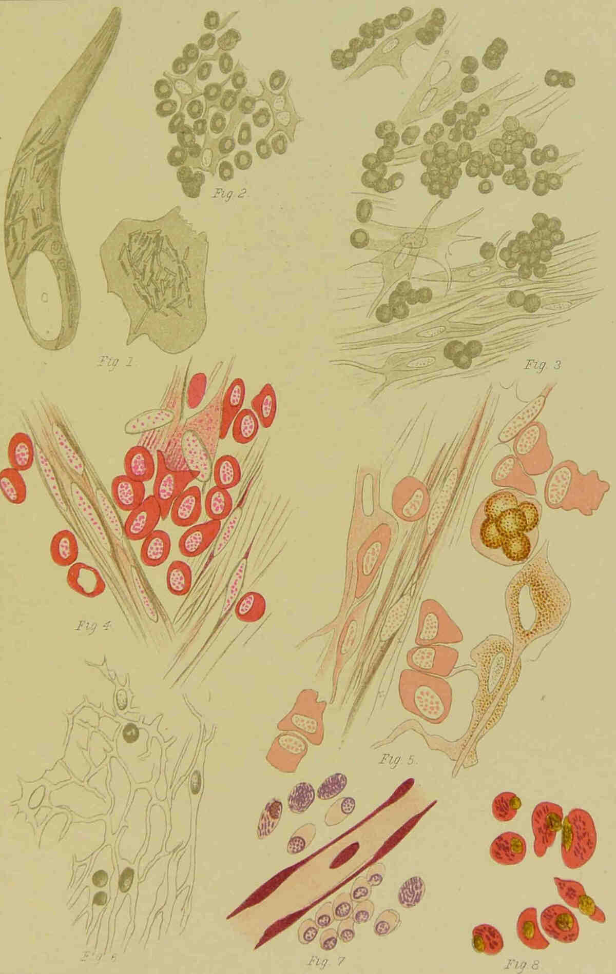
PLATE VII.
PLATE VII.
Fig. 1.—Cells and globi from cutaneous lepromata. Müller’s Fluid (Hartnack, No. ix).
Fig. 2.—Globi from a leprous spleen.
Fig. 3.—Two globi from the retina.
Plate VII.
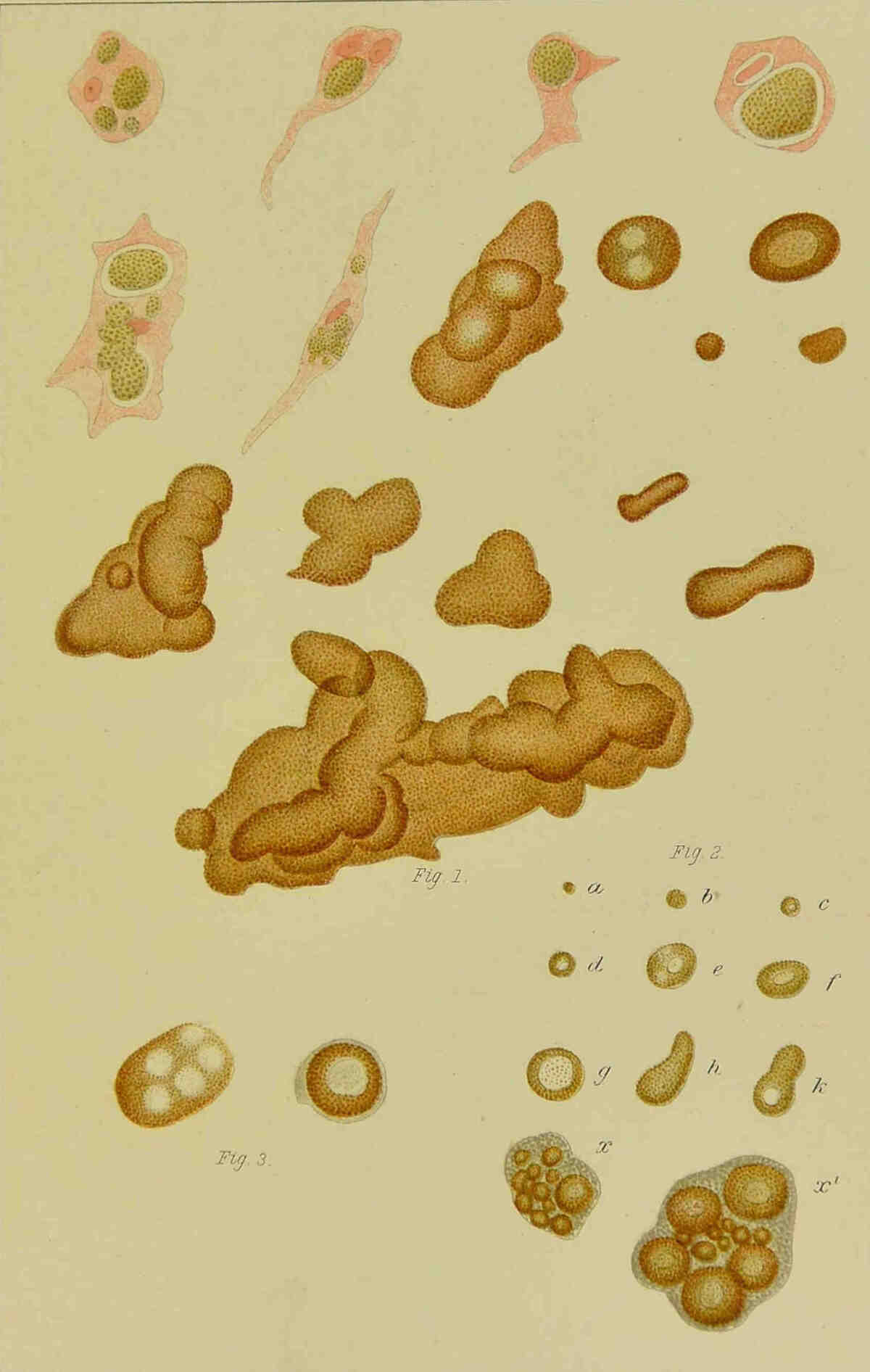
PLATE VIII.
PLATE VIII.
Fig. 1.—Corneal corpuscles filled with brown granules (Hartnack, No. ix).
Fig. 2.—From a leproma of the iris. Round cells with stellate cells (Hartnack, No. ix).
Fig. 3.—Corneal space with round cells near the corneal corpuscles (Hartnack, No. ix).
Fig. 4.—Cells with two nuclei full of bacilli from a cutaneous leproma. Seibert homog. immersion 1/16.
Fig. 5.—Two cells with bacilli from a leprous spleen. Fuchsin, methyl blue. Seibert homog. immersion 1/16.
Fig. 6.—From a leprous liver. White blood corpuscles with bacilli in a capillary. Seibert homog. immersion 1/16.
Plate VIII.
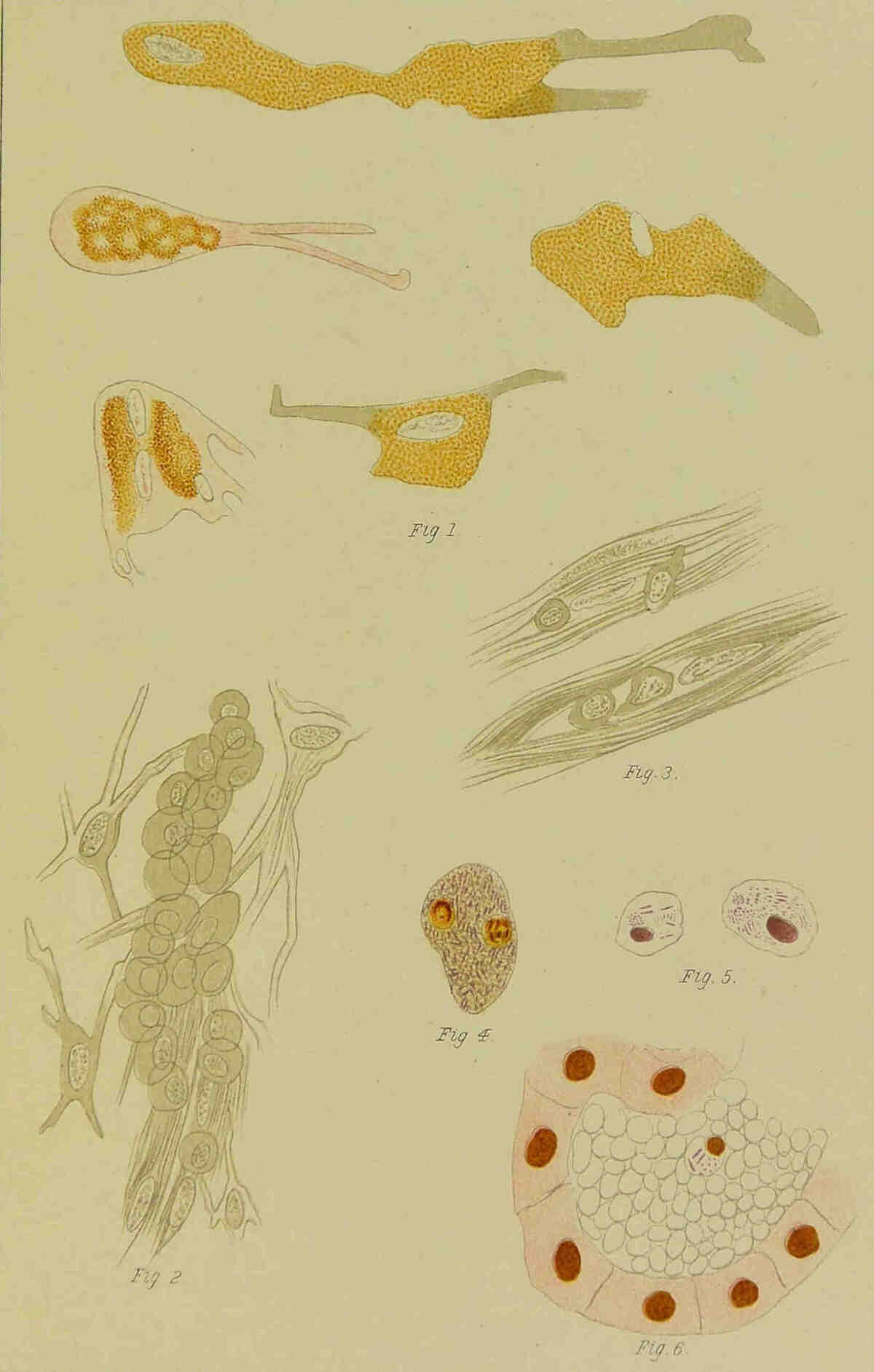
PLATE IX.
PLATE IX.
Fig. 1.—From a leprous liver. Bacilli in an endothelial cell of a capillary. Seibert homog. immersion 1/16.
Fig. 2.—Piece of a very leprous liver. Capsule below, cut surface above.
Fig. 3.—Cut surface of a very leprous spleen.
Figs. 4 and 5 and Plate X, Fig. 1.—Cross section of the seminal canals with bacilli around the nuclei of the walls and in the epithelium; in Plate X, Fig. 1, is shown an epithelial cell filled with bacilli.
Plate IX.
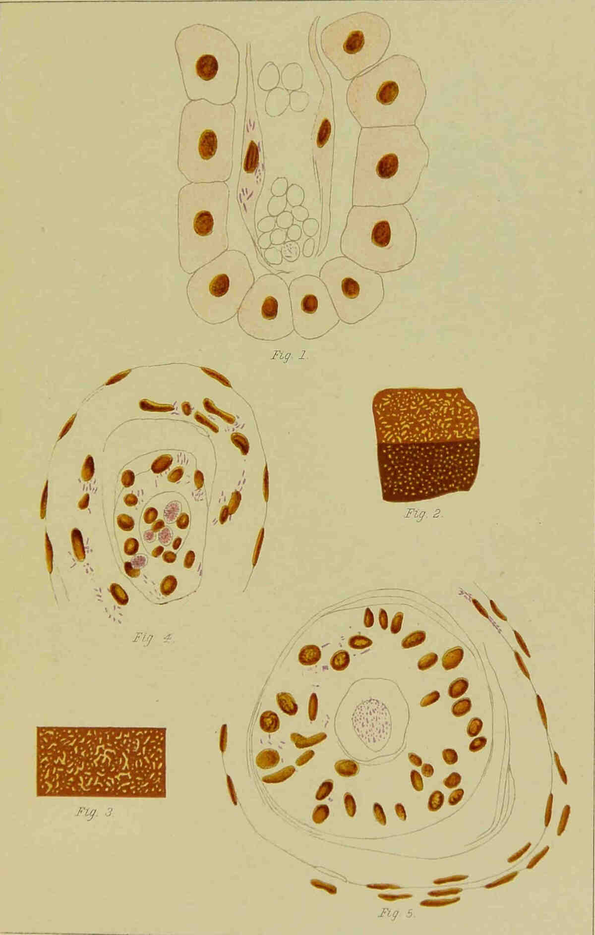
PLATE X.
PLATE X.
Fig. 1.—Epithelial cell from a seminal canal, filled with bacilli.
Fig. 2.—A globus with a vacuole in which are fragments stained with Bismarck brown, probably the remains of nuclei.
Fig. 3.—A globus lying in a cell; the nucleus and a part of the cell-protoplasm preserved.
Fig. 4.—An epithelial cell from a seminal canal, filled with bacilli broken down into granules.
Fig. 5.—Cross section of a blood-vessel with bacilli in the endothelium and a white blood corpuscle filled with bacilli.
Fig. 6.—Longitudinal section of a blood-vessel showing bacilli in the endothelium, and a fibrinous coagulum enclosing two white blood corpuscles, one of them filled with bacilli.
Fig. 7.—Bacilli free between the red blood corpuscles.
Fig. 8.—A connective tissue space from the tunica albuginea filled with bacilli.
Fig. 9.—Cross section of a blood-vessel with bacilli around the nuclei of the surrounding connective tissue.
All the preparations from the testicle were hardened in Fleming’s or Müller’s solutions, and the drawings made with a Seibert homog. immersion lens 1/16. Plate IX, Fig. 5 with Seibert 1/8.
Plate X.
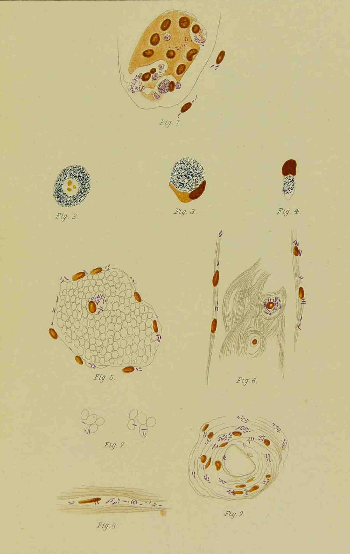
PLATE XI.
PLATE XI.
Fig. 1.—Bundle of nerve fibres from a ciliary nerve, the cells of Schwann’s sheath filled with brown granules.
Fig. 2.—The myelin sheath pressed in by a cell filled with granules.
Fig. 3.—Section from an old leprous macule.
Fig. 4.—Section from a recent leprous macule.
Both sections stained with Fuchsin and Methyl-green. Seibert homogen. immersion 1/16.
Plate XI.
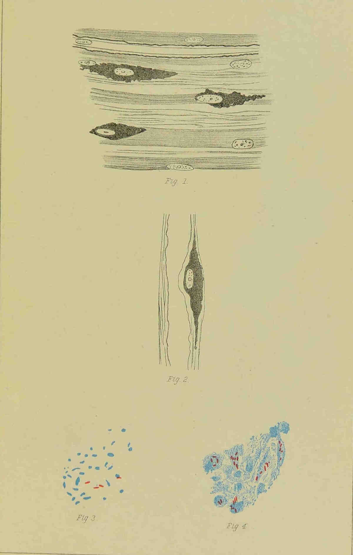
PLATE XII.
PLATE XII.
Fig. 1.—Cross section of a leprous ulnar nerve. The darkly hatched cells represent those filled with brown granules. It will be noted that the axial cylinder is wanting in many of the nerve fibres (Hartnack, No. ix).
Fig. 2.—A piece of neurilemma with flat connective tissue cells lying on it, containing brown granules darkened by osmic acid (Gundlach, No. viii).
Plate XII.
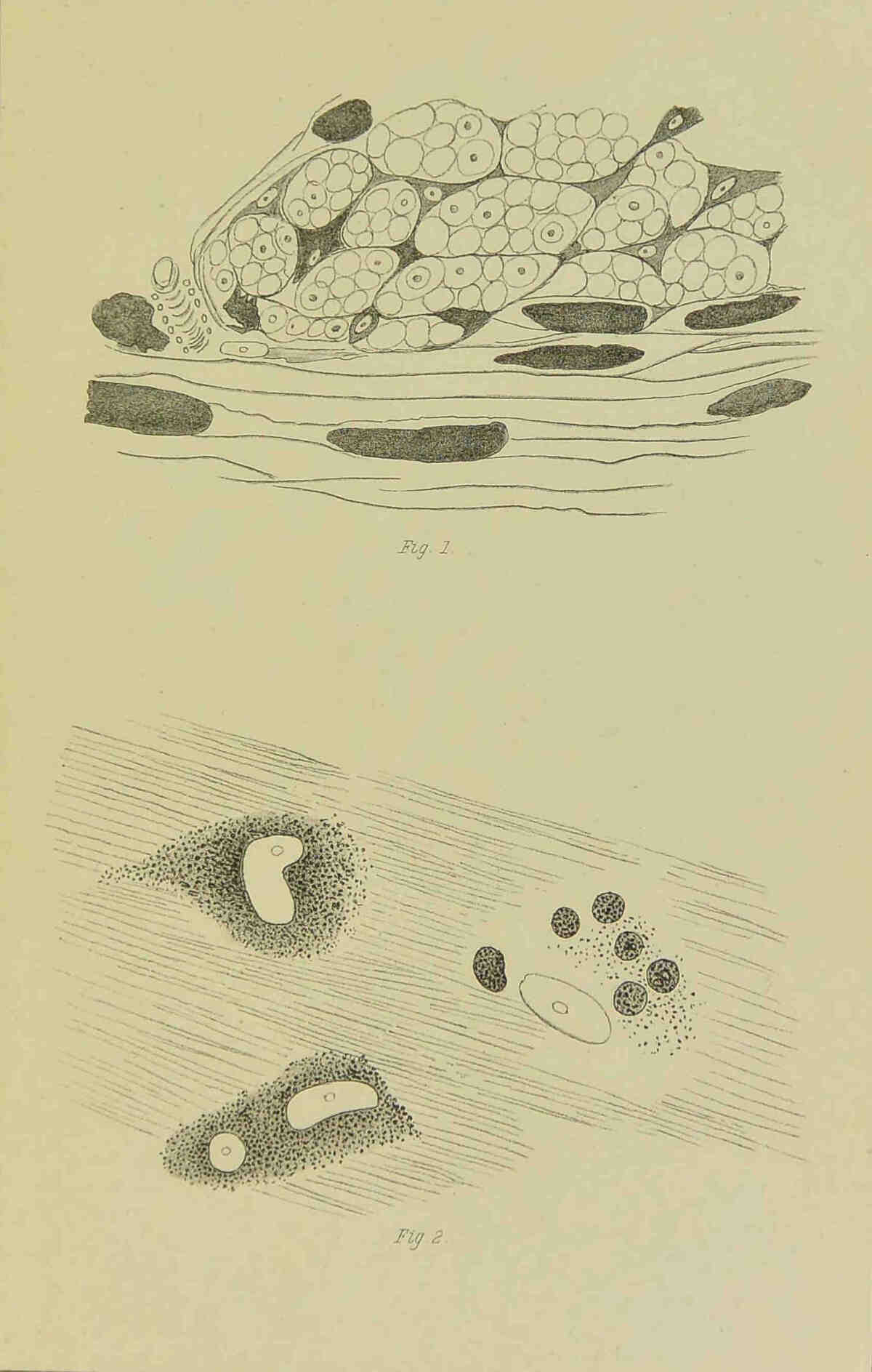
PLATE XIII.
PLATE XIII.
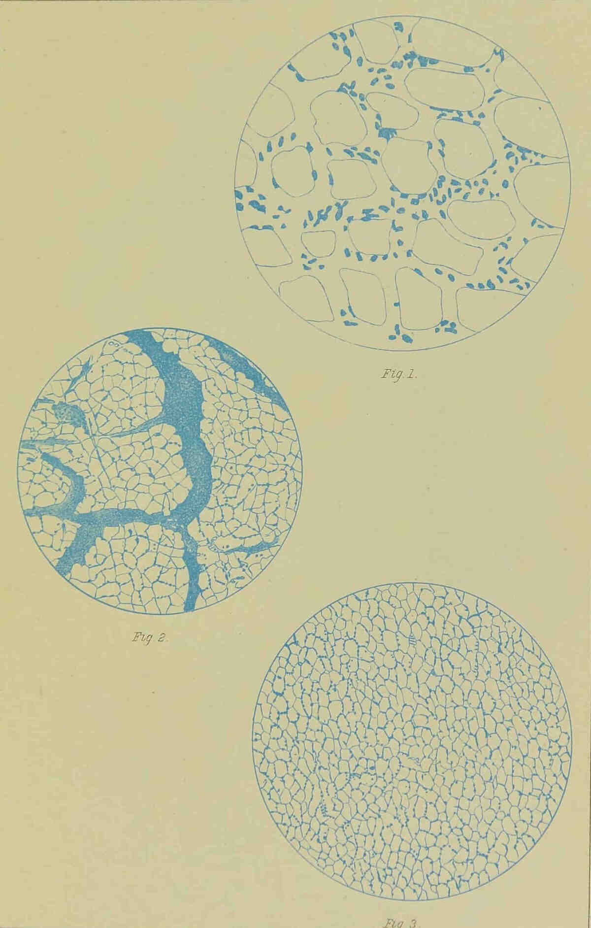
Fig. 1.—Cross section of an atrophic muscle, growth of nuclei in the perimysium. Low power.
Fig. 2.—Cross section of an atrophic muscle with great development of connective tissue. Low power.
Fig. 3.—Proliferation of the nuclei of the perimysium. High power.
Plate XIII.
Transcriber’s Note:
Hyphenation has been standardized.