Chapter IX · (continued 2)
Reading edition
The use of the word 'fungoid,' and particularly that of 'inoculate,' is suggestive enough, and is evidence sufficient that either Blaine or his editor recognised, simply through clinical observation, the working of a special cause.
Four years later, Bouley is found holding the opinion that canker was closely allied to tetter, thus recognising for it a local specific cause. The same observer also pointed out that the secretion of the keratogenous membrane instead of being suspended was greatly increased, taking care to explain, as did Dupuy, that the products of the secretion were perverted and had lost their normal ability to become transformed into compact horn.
In 1864 this slowly growing recognition of a specific cause received further impetus from the statements of Megnier. This observer claimed to have discovered in the cankerous secretions the existence of a vegetable parasite (namely, a cryptogam, as in favus), which he termed the keraphyton, or parasitic plant of the horn.
Modern research, though failing to substitute anything more definite, has not confirmed this. The exact and exciting cause of canker is therefore still an open question, and a matter for research. We may, however, sum the matter up by briefly discussing the causes, so far as clinical observation teaches us. This we shall do under two headings—namely, Predisposing and Exciting.
Predisposing Causes.—Starting with the assumption that the disease is due to local infection, we may relate as predisposing causes anything having a prejudicial effect upon the horn, disintegrating it, and so laying the tissues beneath open to attack. The most prominent in this connection is certainly a continued dampness of the material on which the animal has to stand. Particularly is this the case when the material is also excessively foul and dirty, contaminated with the animal discharges, and presumably swarming with the lower forms of animal and plant life. We shall therefore find bad cases of canker in stables where the "sets" are irregular, or where no paving at all is attempted, where the drainage is defective, and where darkness and want of proper ventilation favours organismal growth. The fact that with modern drainage and a general hygienic improvement in stabling, canker has to a large extent died out, supports this contention.
Again, as with thrush, anything removing the counter-pressure of the frog with the ground and throwing that organ out of play, may be looked upon as a predisposing cause. The atrophy of the frog thus occurring, the deterioration in the quality of its horn and the fissures in its surface lay it specially open to infection. That one of the principal factors in the treatment of canker is a restoration of ground-pressure to the frog and the sole is sufficient proof of this.
Further, it is well to note that, although playing no part in the actual causation, certain constitutional conditions may in some measure predispose the foot to attack. Clinical observation teaches us that animals of a lymphatic nature, with thick skins and an abundance of hair, with flat feet and thick, fleshy frogs, are far more liable to attack than are animals with reverse points.
Exciting Causes. Those who give this subject careful consideration cannot fail to arrive at the conclusion that canker is most certainly due to local infection with a specific poison, and that poison a germicidal one from the ground. The symptoms arising may be due to the action of a single germ, or to two or more germs acting in conjunction. As to whether the parasitic invasion is single or multiple we cannot feel certain, but that it is parasitic we feel absolutely assured.
It is simply the light that bacteriological advance has made during the last two decades that enables us to make the statement with such feelings of assurance. We arrive at our conclusions by reasoning from analogy. Here we have a disease always exhibiting the same symptoms, more or less peculiar to one class of animal, always with a similar characteristic appearance and smell, always obstinately refractory to treatment, showing always a tendency to spread to the other feet of the same animal, and often to the feet of other animals near enough to become infected, and always cured—when cured it is—by a treatment which may be summed up in two words as 'rigid antisepsis.' Other diseases, with points in common with this, have been directly proved to be due to a specific cause. Common regard for logic compels us to admit the same for canker.
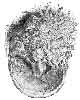
FIG. 134.—A FOOT, THE SUBJECT OF CANKER, SHOWING DESTRUCTION OF THE HORNY FROG, AND A FUNGOID-LOOKING HYPERTROPHY OF THE TISSUES BENEATH.
Symptoms and Pathological Anatomy.—The symptoms of canker are seldom noticeable at the commencement of an attack. The disease is slow in its progress; for some time confines its ravages to the sub-horny tissues unseen, and is quite unattended with pain. It is not observed, therefore, until considerable damage has been done, and the disease is far advanced. What is usually first seen is a peculiar softening and raising of the horn of the frog. The infective material has set up a chronic inflammation of the keratogenous membrane, leading to abnormal secretion, and, in place of the horny cells it should normally secrete, is thrown out an abundance of a serous fluid.
This upraised and softened horn once thrown off is not again renewed, and the whole of the sensitive frog and perhaps a portion of the sensitive sole is left uncovered. In place of the normal horn, however, is often found a hypertrophy of the elements of the keratogenous membrane leading to huge fungoid-looking growths with a papillomatous aspect, damp in appearance and offensive in smell, and readily bleeding when injured (see Fig. 131).
The horn immediately surrounding the lesion is loose and non-adherent to the sensitive structures. This indicates, of course, that the disease has spread further beneath the horny covering than is at first sight apparent. Portions of this loose horn removed reveal beneath it a caseous foetid matter, easily removed by scraping (the perverted secretion of the keratogenous membrane). When this is carefully scraped away, the sensitive structures appear to be covered with a thin, smooth membrane, gray in colour and almost transparent, while beneath it may be seen the red appearance of normal sensitive structures.
If the horn surrounding the lesion is not touched with the knife, but little is seen of the extent of the disease, for that removed by natural means is often very small in quantity. To all intents and purposes the disease appears to be confined to the frog. This appearance is misleading, especially if the disease has been in existence for some time, for it may have easily spread to the whole of the sole, and even to the greater portions of the laminæ secreting the wall.
It is, in fact, not until the pressure exerted by the normal horn is removed by its breaking away that the vascular structures of the keratogenous membrane begin to swell, and the perverted secretions to enlarge in size. Once the pressure is removed, however, this quickly comes about, and the characteristic fungoid growths rapidly make their appearance.
This tendency to spread is highly indicative of canker. The serous matter exuding from the diseased keratogenous membrane appears, in fact, to be highly infective. Once its flow is commenced, it slowly, but surely, invades the sensitive structures near it, appearing, as Elaine has put it, to 'inoculate' them. What is really the case, of course, is not that the discharge itself is infective, but that it is contaminated with infective material.
The fungoid-looking growths to which we have before referred are, in reality, nothing more than the villi of the sensitive frog and sole greatly hypertrophied and irregular in shape. At times the hypertrophy is as a huge and compact enlargement occupying the position of the frog. Sometimes, however, it occurs as numerous elongated and twisted fibrous bundles, separated from each other by deep clefts, and the clefts filled with the offensive cankerous discharge (see Fig. 134).

FIG. 135.—LOWER ASPECT OF CANKERED FOOT, SHOWING DESTRUCTION OF WALL.
At a very advanced stage canker leads to destruction of much of the horny sole and frog; or even parts of the wall may become separated from the tissues beneath, and break away from the foot (see Fig. 135). At other times the disease brings about a deformity of the whole of the foot. Its longitudinal and transverse diameters become enormously increased, and the whole foot apparently flattened from above to below (see Fig. 136). This indicates that not only has the horny sole been entirely destroyed, but that the destructive process has also extended to the greater part of the lower half of the wall, with a consequent hypertrophy of exposed soft structures, and a sinking of the bony column, similar to that which occurs in laminitis, but not so pronounced.
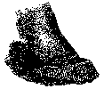
FIG. 136.—FOOT WITH ADVANCED CANKER.
A further aspect of the badly-cankered foot is to be found in an apparently enormous increase in the length of the wall. This we have seen protruding for quite 5 inches beyond the plane of the sole. It simply indicates that, in order to keep the animal at work, the smith has at every shoeing spared the wall, so that the diseased structures might be kept from contact with the ground.
As we have said before, pain and other symptoms of distress are quite absent. Animals affected with canker for a long time maintain their condition, feed well, and are quite capable of performing work under ordinary conditions.
Differential Diagnosis and Prognosis.—Perhaps the only disease with which canker may be confounded is thrush. They should, however, be easily distinguishable. The discharge from thrush is not so profuse, and is thicker and darker in colour, while the loosening of the horn is almost entirely absent. Furthermore, thrush shows no tendency to spread, and, even when left untreated, may remain confined to the frog for months, and even years. Canker, on the other hand, is slowly progressive, and soon shows the characteristic fungoid excresences, which growths are in thrush never seen. A further point of difference is discovered when treatment is commenced. Canker is found to be refractory to a point that is absolutely disheartening, while thrush, with careful attention, is soon got under hand, and a permanent cure effected.
The prognosis must be guarded. By many canker has been said to be incurable. This, however, has been clearly shown to be wrong. When the animal is young, and treatment may reasonably be judged to be economical, then a favourable prognosis may be indulged in, provided the veterinary surgeon intends to put into that treatment a more than ordinary amount of individual care and attendance. Even then, however, he will have to be very largely guided by the condition of his case. He should see that it is not too far advanced, and that a great deformity of the hoof, or actual exploration, does not indicate disease of the greater part of the wall.
Treatment.—From what has gone before, it will be seen that the eradication of canker is no easy task, that it is, in fact, a most difficult matter, and one not to be lightly undertaken. At the risk of recapitulating what we have said before, we may mention here the two points which the veterinarian must bear in mind. (1) That there is no actual disease or alteration in structure of the deep layers of the keratogenous apparatus. It is only the superficial, or horn-secreting, layer that concerns us. (2) That the disease of this superficial layer is infection with a material that may reasonably be presumed to be infective.
Put thus, treatment of canker would at first sight appear to be easy. One would imagine that a simple and long-continued soaking of the entire foot in a strong enough antiseptic would be all that was needed. Clinical observation, however, shows that this is not so, and for this there must be reasons.
The reasons are these: (1) Between us and the diseased layer upon which our attention must be directed is often a layer of normal horn, effectually protecting the tissues beneath from any dressing which we might consider beneficial. (2) Anything applied with the object of destroying septic material, but strong enough, or caustic enough, to injure the membrane upon which we are working, only makes the case worse. The superficial layer of the keratogenous membrane in which we have judged the disease to exist is, after all, but a delicate structure. When attacked by the application of too potent a drug its horn-secreting layer is easily destroyed, and thus, although we may succeed in establishing asepsis, we cannot expect at the point of injury a growth of horn. In its place we are confronted with large outgrowths of inflammatory fibrous tissue. (3) Shedding of the diseased horn and removal of the pressure exerted by the hoof faces us with hypertrophy of the exposed villi. The difficulty of meeting this with an adequate and evenly-distributed pressure is well enough known, and we find in that a further reason that the treatment of canker is superlatively difficult. (4) The material on which the animal has to stand is a distinct bar to the maintaining of a strict asepsis.
When we have said this, it is easy to understand that canker is not to be successfully met with any so-called specific—that it makes but little difference what the application may be so long as it is antiseptic, and is used by a man thoroughly conversant with the difficulties he has to contend with, and with his mind firmly set upon surmounting them.
With this point established, we will not devote more of our space to a consideration of the various dressings that have at different times been highly advocated in the treatment of the disease. It is interesting, however, to note that intensely irritating and caustic applications have been greatly in favour. Nitric acid, sulphuric acid (either alone or its action reduced by the addition of alcohol, oil, or turpentine), arsenic, butter of antimony, creasote, chromic acid, carbolic acid, arsenite of soda, and the actual cautery, have all been used.
Without dwelling further on that, we may say at once that a correct treatment consists in (1) the removal of all horn overlying infected portions of the keratogenous membrane, (2) the application of an antiseptic not too powerfully caustic in its action, (3) frequent changes of the dressings in order to insure a maintenance of antisepsis, and (4) the application of an adequate pressure to the exposed soft structures. Thus combated, canker is curable.
The man who, at the expense of much time and trouble, has demonstrated the truth of these axioms is Mr. Malcolm, of Birmingham. The determination with which he clung to his point that canker was, with correct treatment, in every case curable, was some years ago provocative of much discussion in veterinary circles. That he was successful in proving his contention is more to our point here. It is his method of treatment, therefore, that we shall give, and this we shall do by liberal extracts from Mr. Malcolm's own writings.
'On the first occasion of operating upon and dressing the cankered foot, it is usually necessary to cast the horse, and this may have to be done at intervals for a second or even third time; but in most cases once is sufficient, subsequent dressing being usually accomplished without much difficulty, frequently even without the aid of a twitch. After the horse has been secured, the drawing-knife is first employed; and if the frog alone is affected, it is unnecessary even to pare the sole, the removal of all frog horn not intimately adherent to its secreting surface being all that is required. But if both sole and frog be involved, the whole of the sound horn should be first thinned until it springs under the thumb, and then, using a sharp knife, every particle of diseased horn must be carefully removed from both sole and frog, a process much more easily, and with far greater certainty, secured by the previous thinning of the horn.
'The removal of diseased horn should always commence at the most dependent part of the foot, so that any hæmorrhage produced may be below the parts still to be operated on, a matter of considerable moment for effective treatment. But with due care there will be little hæmorrhage, as, except in the initial stage, there is no real union between the diseased horn and the diseased vascular secreting surface.
'After all apparently diseased horn has been removed by the knife, any still remaining should be at once destroyed by the actual cautery, by which it can be identified. All the diseased secreting surface should be carefully scraped with a thin hot iron,[A] fungoid growths excised and cauterized, and, indeed, every particle of cankered tissue should, if possible, be eradicated. In securing this more reliance can be placed on the actual cautery than on any other, whether liquid or solid: it is more under control in application, more decisive in effect, and its results can be anticipated with a far greater certainty. Moreover, its aid in diagnosis is of immense value; applied to the thinned horn or secreting surface it unmistakably demonstrates the presence or absence of canker. Healthy tissue chars black; cankered tissue, on the contrary, bubbles up white under the hot iron, and presents an appearance not unlike roasted cheese.
[Footnote A: The words in italics are alterations in the original article made by Mr. Malcolm in a private letter to the author (H.C.R.).]
'Although this test is certain for horn thinned to the quick, it is not to be relied upon with thick horn, the outside of which may be practically healthy and char black, while its underlying surface may be cankered. With this exception the test is an infallible one, as by it the demarcation between cankered and healthy tissue can be clearly traced, and as a result we can with equal confidence radically remove[A] all cankered tissue, and conserve all healthy. As the object of that abominably cruel and barbarous operation of stripping the sole is the exposure of all canker, and as this can be done with equal certainty with the aid of the hot iron, there can be no necessity for performing it. The pain of cauterizing cankered tissue, which is a necessary operation, is infinitesimal (canker largely destroying sensation), compared with the pain produced in the totally unnecessary process of tearing healthy horn from a highly sensitive tissue.
[Footnote A: The words in italics are alterations in the original article made by Mr. Malcolm in a private letter to the author (H.C.R.).]
'Having by means of the knife and cautery removed every known particle of disease, the next procedure is to pack the surface of the sole and frog thus exposed with a mild dressing, such as vaseline; but if the cankered surface has not been efficiently, scraped, than there is required a more [A] powerful astringent or caustic dressing, which may vary considerably according to the individual fancy. A great favourite of mine consists of equal parts of sulphates of copper, iron, and zinc, mixed with strong carbolic acid, a very little vaseline being added to give the mass cohesion. The dressing, covered by a pledget of tow, is held in position by a shoe with an iron or leather sole, and the dressing and tow together should be of sufficient bulk to produce slight pressure on the sole when the nails of the shoe are drawn up. This insures contact between the dressing and the exposed surface, as well as any benefit derivable from pressure.
[Footnote A: The words in italics are alterations in the original article made by Mr. Malcolm in a private letter to the author (H.C.R.).]
'The dressing of the foot and nailing of the shoe can usually be more expeditiously performed when the horse is on his feet than when prone. If only the frog, or the frog and a small part of the sole, be involved, the horse should be kept at work, but if a large part or the whole of the sole a few days' rest may be necessary; but as soon as the condition of the foot will allow, work should be resumed, and it is simply marvellous how sound a horse will walk while minus the greater part of his sole from canker.
'On the second day following the shoe should be removed, and the foot redressed. To effect this it is necessary to recast the horse. Commencing at the edge of the sound horn, at the most dependent part of the foot, all new horn, no matter what its condition, must be pared to the quick, especial care being taken to effectually remove any lingering disease. Want of success is frequently attributable to neglect of this precaution. A small particle of canker remains undetected, forms a new centre of infection, and just when success is anticipated, much to your chagrin you have to deal with a fresh outbreak of canker, instead of a rapidly-healing foot. Parenthetically, I may here remark that the amount of more or less imperfect new horn produced by a cankered surface after an effective but not too destructive cauterization is almost incredible, and one cannot fail to be struck with the very active proliferation here compared with the meagre production of new horn by the healthy surface.
'After all disease has been excised, carefully clean the foot with waste, thoroughly protect any raw surface resulting from overcauterization by some mild agent, such as a saturated calomel ointment, reapply an astringent dressing over the whole affected surface, and nail on the shoe. This method of procedure should now be thoroughly carried out daily for a time, and as it is proceeded with a successful issue soon becomes assured in nearly every case. Where, in spite of these efforts, the disease still persists, depend upon it the fault is with the operator, who has failed to eradicate some centre of infection. Under these circumstances it may be necessary to recast the patient, repare the foot, and by the aid of eye, knife, and cautery, endeavour to find the cause, and having found it, which can invariably be done, remove it. The usual treatment will then speedily become successful. As the case proceeds dressing every other day will soon be sufficient, then twice a week, and finally, once a week until sufficiently cured.
'During this healing process, and after the complete eradication of canker it may be again repeated, no agent seems to have a more beneficial effect than calomel, and for this purpose it is best used as a dry powder. Under this dressing any remaining spot of canker is readily detected by the wet condition of the calomel when the shoe is removed the next day. In dealing with such a spot, a very good plan, after all apparently diseased tissue has been excised, is to touch the cankered part with solid nitrate of silver, or a feather dipped in one of the strong mineral acids, and then reapply calomel over the surface. The result of this treatment is frequently very gratifying.
'In successful treatment the shoe must be removed each time—an adjustable plate will not do, as no man can thoroughly pare and examine a foot with the shoe on, and imperfect dressings are worse than useless. Indeed, it is better not to pare or thin the horn at all, than to imperfectly pare, since canker, if undestroyed, develops far more rapidly under thin horn than under thick.
'In conclusion, I would again urge the necessity, at the very first operation, when the horse is down, of removing every single particle of the diseased tissue, either by excision or effectual cauterization, but at the same time taking very great care to guard against the latter being too destructive. The cautery should be laid aside as soon as the tissue cauterized ceases to burn white. The moment at which the canker has thus been eradicated without destroying sound tissue is indicated by the appearance of healthy horn, by the intimate union of that with the secreting surface, and by the healthy aspect of the exuded blood when paring has been carried to the quick.
'Should subjacent healthy structures be destroyed during the process, that is shown by the production of a raw sore, or of a sore to which a "sit-fast," coextensive to the injury, is firmly attached. This seriously retards recovery. The secreting surface having been destroyed, no new horn can be produced directly from the part, and a new secreting surface and new horn have now to grow inwards from the surrounding undestroyed tissue, and that is a slow process. At the same time, on the principle of choosing the least of two evils, practical experience teaches that it is better to produce a small sore or a "sit-fast" than to leave a part of the canker undetected; but, on the other hand, it is better to leave a small part of canker undetected, which can be recognised and removed at the next examination, than to cause a large slough. The object of the skilful surgeon is, naturally, to avoid both extremes; and if trouble be taken to carry out the procedure described, there need be no fear of the result.'[A]
[Footnote A: Journal of Comparative Pathology and Therapeutics, vol. iv., p. 24.]
Treated in this way, the horse with cankered feet may be usually kept at work during the whole time that treatment is carried out, and a cure is obtainable in periods varying from six weeks to six or even twelve months.
The same essentials in treatment—namely, removal of diseased horn, antiseptic dressings, and pressure—are insisted on by other writers. Bermbach,[A] in 1888, treats canker as follows: The horse having been cast, the undermined hoof-horn is removed with the knife, and the hypertrophied sensitive structures, if necessary, reduced in the same manner. The chief difficulty in removing the latter is experienced in the lateral lacunæ of the frog, where it is most conveniently scraped away with a spoon or sharp curette. Professors Hoffmann and Imminger also operate in the same way, applying an Esmarch's hæmostatic bandage, and using the knife and curette freely.[B]
[Footnote A: Ibid., vol. ii., p. 68.]
[Footnote B: Veterinary Journal, vol. xxxv., p. 433.]
Hæmorrhage is afterwards arrested, and a dressing of perchloride of mercury (a solution, 1/2 per cent., in equal parts of alcohol and water) applied. The after-dressings succeeding best are those of slightly caustic and astringent agents, preferably in the form of a powder, and held in position by carbol-jute pads and linen bandages applied with a certain amount of pressure.
The same author draws attention to the fact that caustic agents such as nitrate of lead, chloride of zinc, etc., act too powerfully if the bleeding has been arrested and the wound disinfected. They then form a thick crust, under which profuse suppuration takes place. The same agents are likewise contra-indicated when hæmorrhage is still present. In this latter case they combine with the blood to form metallic albuminates, which lie as an impenetrable layer on the surface of the wound, and so hinder the action of drugs on the tissue below.
During his after-treatment, Bermbach advocates removal of the dressings every second day, all cheesy material to be scraped away with the knife, and the sublimate lotion to be used again. He also insists on the animal being kept standing in a dry stable,—nothing but a stone pavement kept clean—and put to regular work in a plate shoe after the first or second week. Cure of advanced cases is said to be obtainable in from four to six weeks.
As illustrative of the value of pressure in the treatment of canker, we may also draw attention to a treatment advocated by Lieutenant Rose.[A] This observer holds that adequate pressure is unobtainable by packing the foot, and, to obtain it, removes the wall from heel to heel, much after the manner of preparing the foot for the Charlier shoe, so that the whole of the weight is taken by the sole and the frog. Tar and tow is then lightly applied, the foot placed in a boot, and the patient turned into a loose-box. The dressing is repeated at intervals of four or five days until the animal is cured.
[Footnote A: Veterinary Record, vol. xi., p. 435.]
Those who have followed this method of treatment have modified it by actually shoeing the animal Charlier fashion, and keeping him at work, attention, of course, being at the same time given to a proper antiseptic dressing.
Reported Cases.—1. (Malcolm's Treatment[A]). The subject was a five-year old horse belonging to a client of Mr. Giver's, of Tamworth. The case was an exceptionally bad one, for not only was the whole of the frog and sole of the near hind-foot cankered, but the disease on the outside quarter extended to within 1/2 inch of the coronet, and on the inside quarter to within 2 inches of it. As the owner, a farmer, had not proper convenience for Mr. Olver to treat the case, the latter asked me, while visiting him, if I would care to undertake the treatment, saying at the time it would be a very good test-case, as the disease was so far advanced. I readily agreed, and, after the necessary arrangements, had the horse removed to Birmingham on July 2. In this case it was found necessary to cast the animal and cauterize the foot a second time before a healthy granulating surface was secured; but after this the progress towards recovery was uninterrupted, although necessarily slow, on account of the large amount of new secreting surface which had to be formed.
[Footnote A: Journal of Comparative Pathology and Therapeutics, vol. v., p. 48.]
The horse was finally discharged, after inspection by Mr. Olver, absolutely cured and free from canker, on January 7.
The illustration (Fig. 135, p. 312) is from a photograph, and it gives a somewhat imperfect representation of the state of the foot two months after it came under my care.
2. (Rose's Treatment.[A]) This was a bad case of canker, which had been for two or three months treated in the ordinary manner, with but little sign of ultimate success. Commenced in June and carried on until the end of September, the ordinary treatment consisted in burning down the fungus growth with the hot iron, and dressing with copper sulphate, zinc sulphate, and boracic acid. The cauterization was repeated every five days.
[Footnote A: Veterinary Record, vol. xi., p. 435.]
The treatment of Lieutenant Rose was commenced at about the end of September, at which date the disease extended from the toe on one side of the foot right back to the heel, involving the sole, half of the frog, and the bulb of the heel. One week after treatment the diseased surface was drier, and granulations were more healthy. At the expiration of a fortnight the new horn had commenced to grow from the wall, and also from the frog, right round the diseased surface, the diseased part of the bulb of the heel being divided from the sole by new horn.
Three to four weeks later the diseased surface was gradually getting smaller, while in about six weeks it was quite healed up, the last place to heal being a strip outside the bar, between it and the wall, and a smaller spot on the bulb of the heel. These healed up simultaneously, and left the animal sound.
3. (Treatment by Pressure, H. Leeney [A]). I was consulted in the early part of last summer, before the dry weather had begun, as to a farm-horse with canker in three feet. Her shoes were in the 'disgruntle' condition we so often find on farms, that, to give her a level bearing until I should call another day with a farrier to help me to pack the foot up in the old-fashioned way, I had the remaining shoes pulled off. The case somehow dropped out of my list, and I neglected to call, until asked one day to see something else.
[Footnote A: Veterinary Records, vol. xi., p. 447]
I then found that, under a pressure of work, the animal had been used in the shafts of a farm-cart on tolerably level ground, and when the dry weather had already set in. There was a distinct improvement in all the diseased feet, and as she was badly wanted I contented myself with rasping off some broken crust, and supplied some caustic dressing for use at night. Without shoes she worked continuously on the dry and hard meadow-land for several weeks, and was practically cured in something less than three months. My astringent or caustic lotion may have had something to do with the cure of the deep-seated parts, but the bare recital of the case should be sufficient to show that it is all a question of bearing, or nearly so.
7. SPECIFIC CORONITIS.
Definition.—In describing this condition under the above heading, we are following the lead of Mr. Malcolm. We may define it as a chronic inflammatory condition of the keratogenous membrane, usually confined to that of the coronary cushion, the ergots and the chestnuts, but sometimes extending to that of the frog and the sole, characterized by a malsecretion of the affected membrane similar to that observed in canker.
Causes.—The cause which we have indicated for canker—namely, a local specific one, is in all probability the one operating here. Apparently there is a variance of opinion as to whether the condition is actually canker or not. We think, however, that the character of the secretion of the affected membranes, the appearance of the growths, the manner in which they react to the hot iron, the comparative absence of pain, and other points of similarity, point to the fact that the two conditions are actually identical. In other words, the cause is precisely the same, and the only point of difference is the alteration in the point of attack.
Symptoms.—Like canker, the disease is insidious in onset. In precisely similar manner the horn, and in this case the skin of the coronet, is underrun. Later there is the partial shedding and fissuring of the undermined horn and the exuding of the characteristic discharge—in this case not so watery as that of canker. The caseous material of canker is also present, as is a disposition to hypertrophy of the exposed sensitive structures. What horn is left becomes rough and irregularly fissured, and has been likened by some observers to deeply-wrinkled bark of an old tree. A peculiar characteristic of this condition is the state of the ergots and chestnuts. Here the keratogenous membrane participates in the diseased process, and their horn becomes dry and brittle, and readily splits into small fibrous bundles very similar to the fibroid growth described in canker. These excrescences are easily separated from the sensitive structures beneath, and the exposed surface is seen to be more or less moist, or even exhibiting a slight oozing of blood.
Again, as in canker, the deeper layers of the sensitive structures appear to be normal, the horn-secreting layers being the only ones affected. According to Malcolm, the disease is in its nature equally as inveterate as canker, but it is easier to treat, on account of its more exposed position.
Treatment.—This is exactly that as described for canker.
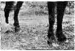
FIG. 137.—SPECIFIC CORONITIS OF ALL FOUR FEET.
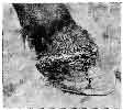
FIG. 138.—OFF FORE-FOOT AFFECTED WITH SPECIFIC CORONITIS.
Recorded Case.—The subject of this case was a young black cart gelding. The disease is reported as having begun as thrush, and then extended to the coronet. When I saw him he had been in a similar condition to that depicted in Fig. 137 for, it was said, two or three months, the driver of the horse meanwhile endeavouring to effect a cure by some potent drug of his own. The animal was in good condition, but walked with difficulty owing to the pain. The coronary bands were swollen to two or three times their natural size, and this caused the hair immediately above to curl upwards. Just below the coronary bands there was a line of separation between them and the wall. They themselves were covered with the cheesy substance typical of canker, and they bled on friction. Down the wall of the off fore-foot some blood had trickled, which may be seen in Fig. 138. The frogs of all four feet bulged backwards, and were badly affected. The soles were covered with normal horn, but I did not resort to paring to see if they were affected. One very curious feature about the case was the fact that all the callosities (ergots and chestnuts) seemed to participate in the morbid process, and they, too, were covered with a thin layer of soft cheesy horn. The animal used to bite at his coronets and also the callosities above the knees and hocks until they bled, which they did quite easily. The owner would not go to the expense of having him treated, so he was destroyed.[A]
[Footnote A: Henry Taylor, Veterinary Record, vol. xvii., p. 311.]