SECTION X. INFECTIOUS DISEASES.
Reading edition
COW-POX—VACCINIA.
The name cow-pox, or vaccinia, is employed to describe a special disease which in animals of the bovine species is characterised by the development of pustules at points where the skin is fine, and more particularly the mammary region.
It can be conveyed both to man and the domestic animals.
This disease has been known from time immemorial, and it would appear that first of all in the East and later in England it was a general belief that its attacks rendered human beings proof against small-pox. Medical men, it must be admitted, long regarded this belief as a popular delusion, as is proved by their continuing to practise inoculation with true small-pox material.
Jenner in 1770 was the first to declare the truth of this popular opinion, and by his wise foresight to confer on humanity one of the most beneficent discoveries ever made, although the weight of modern opinion is in favour of the identity of cow-pox and human variola. Having observed that milkmaids who happened to have small cuts or sores about the hands sometimes contracted the disease in a mild form, and that they did not afterwards suffer from small-pox, he was struck with the advantages consequent on such a discovery, and having proved the possibility of inoculating human beings artificially, he immediately formulated the principles of vaccination. A child eight years of age was vaccinated with cow-pox, and afterwards inoculated with pus from a small-pox patient. It contracted vaccinia in consequence of the first inoculation, but entirely resisted the attempt to inoculate it with small-pox. Vaccination had been discovered.
Jenner furthermore proved that cow-pox was transmissible from cow to cow and from man to man, but it seemed to him that the original disease was to be sought elsewhere, and that the pustular affection originated primarily with the horse. The horse is sometimes the subject of a pustular disease called horse-pox; this disease when inoculated in man confers immunity against small-pox, just as does cow-pox, and Jenner believed that the disease did not attack 666cows unless they had been accidentally inoculated through the medium of the people about the farm. Unfortunately, he named the pustular disease of the horse which he had studied “sore heels,” and for a long time all those who busied themselves with the question of vaccine confounded “sore heels” with a number of different diseases, although as early as 1802 Loy had experimentally proved that so-called “grease” (in reality horse-pox) was transmissible by inoculation to the cow, in which it produced cow-pox.
Loy’s “grease” and Jenner’s “sore heels” only represent forms of horse-pox, but for more than fifty years the origin of vaccine was sought in grease, lymphangitis, and other diseases which attack the extremities of horses’ limbs. Pételard (1845–1868) rediscovered and redescribed horse-pox and proved its transmissibility to man; Lafosse and U. Leblanc discovered it in an epizooty which broke out at Rieumes; and Bouley in 1862 furnished a synthetical description of it under the designation of horse-pox. He shows that horse-pox is always a pustular disease, but that it may sometimes appear in the form of a discrete eruption around the lips and nostrils, sometimes of an eruption limited to the pasterns or extremities of the limbs when inoculation has been effected in this region, sometimes of lymphangitis, and sometimes of a more or less confluent and generalised eruption.
Symptoms. The disease as discovered and described by Jenner was soon rediscovered and redescribed on all sides—by Sacco in Italy, Hering in Germany, etc.
The pustular eruption usually appears on the udder in the case of cows, and on the muzzle, nose, and lips in that of calves. In exceptional cases the eruption may become generalised.
The pustules are round or slightly elliptical, and are preceded by the appearance of red congested patches, followed by infiltration and thickening of the skin.
The pustule is moderately prominent, and after some days there is exudation at its centre, transforming it into a vesico-pustule. The exuded liquid collects under the thickened layer of epidermis, which it raises, and on examination it appears as a white or transparent little central patch, with a thin grey periphery surrounded by a reddish inflammatory zone. This liquid becomes thicker and the pustule is flattened at its centre, then, towards the eighth or ninth day, the pustule is ruptured, owing to tearing of the epidermic patch. The vaccine thus escapes.
In what is termed spontaneous vaccinia the udder is covered with a varying number of pustules, usually in different stages of development. Some are very small, whilst others have attained the size of sixpence and are already in course of cicatrisation.
667When cow-pox is accidental or the result of inoculation, the eruption occurs exactly at the point of inoculation, whether the latter has been through an abrasion, a puncture, an incision, or any other skin injury, and the eruption may assume the most varied appearances, according to the nature of the primary lesion, although the mode in which the pustules themselves form never varies. Pustules experimentally produced by puncturing the parts may be taken as a type of inoculation. On the day following the operation nothing abnormal appears. On the third day there is a slight swelling around the point punctured, and this increases until the fifth day, when there is exudation, which converts the primary lesion into a vesico-pustule. On the sixth day the vesico-pustule becomes umbilicated at its centre, the exudation is abundant, and already vaccine might be collected.
This may be termed the period of crisis; the appearances are most characteristic. During the following days the vesicle is ruptured; the discharge continues from the ninth to the twelfth day, when the pustule diminishes in size and dries up. After the fifteenth day crusts of a brownish colour form; these separate between the twentieth and twenty-fifth days, leaving hard whitish-looking cicatrices, which permanently remain.
Moderate itching accompanies the development of the eruption, the principal functions are not disturbed, and fever only appears in the event of the eruption becoming confluent or extending over a large area. When the eruption is generalised the pustules or vesico-pustules are found mostly in the region of the elbow, the lower border of the neck, the flank and the inner surface of the limbs. They present exactly the same appearance as the pustules on the surface of the udder, but, being covered with hair, are less open to inspection.
In certain rare cases the eruption extends to the perineum and lips of the vulva. Signs of inflammation then develop on one or both sides, the tissues display œdematous infiltration and disseminated or confluent pustules. The lymphatic glands and vessels in the neighbourhood of the pustules are always swollen.
Causation. Cow-pox, or vaccinia, is a virulent disease transmissible by accidental or intentional inoculation. The discharge from the vesico-pustules and the crusts which afterwards cover them are virulent, and inoculation can be performed by simply scratching the skin. A first attack confers prolonged and sometimes perfect immunity, the operation being successful if only one pustule develops.
The disease is transmitted to healthy animals by milkers, by calves in sucking, or by the conveyance in whatsoever form of virulent material to sores or cuts.
The nature of the parasitic or microbic agent which produces the 668disease is still unknown. Some investigators have described intracellular parasites, others extra-cellular parasites, others, again, blood parasites, etc., but the exact cause has always eluded research.
It is, however, known that filtration of vaccine, pure or diluted, through porcelain removes the active material, which remains in the residue arrested by the filter.
Prolonged exposure to a temperature above 104° Fahr. (40° C.) greatly diminishes the activity of the vaccine. Simple desiccation has no action. Warming to 140° Fahr. (60° C.) for fifteen minutes also destroys its action completely.
These facts explain why it has always been so difficult to preserve and cultivate vaccine in tropical regions.
Mixed with equal parts of neutral glycerine, the virulent material preserves its activity unimpaired for from six to eight months.
The disease develops equally in man, the horse, the ox, the buffalo, the goat, and the camel. Its development is less typical in the pig, sheep, dog, and rabbit. Young animals are best adapted for its cultivation.
The blood and serum of animals suffering from cow-pox possess immunising properties, but only when administered in very large doses, say from 6 to 12 lbs. of blood, or ½ to 1 lb. of serum.
The curative action of this serum against small-pox is comparatively trifling.
The diagnosis of cow-pox is not very difficult.
At first the disease might be mistaken for false cow-pox, the eruptions of foot-and-mouth disease, or gangrenous coryza.
In false cow-pox, the nature of which is also little understood, although it is known to be contagious and is regarded by some as true vaccinia, the pustules are smaller and thinner, while the vesicle is more developed, and the disease runs a more rapid course.
In foot-and-mouth disease the eruptions are of the nature of vesicles or bullæ, not pustules. The eruption occurs in twenty-four hours or less, and can only be mistaken for vaccinia during the period of desiccation and the formation of crusts.
Finally, as regards gangrenous coryza, the hardened pustules do not produce vesicles.
The prognosis is generally favourable. The disease runs its course within relatively fixed periods, according to the development of the pustules, and recovery occurs without complications.
Treatment. No curative treatment can be laid down, the development of the disease being perfectly regular and tending to recovery. Simple hygienic precautions and cleanliness are sufficient to avoid complications due to suppuration.
669
COW-POX AND HUMAN VARIOLA—PREPARATION OF VACCINE.
Time and experience having proved that inoculation with cow-pox or vaccinia protected human beings against small-pox, the question arose as to the connection between the two diseases, whether or not they were identical and whether vaccinia in the bovine animal might not merely represent an alternative form of small-pox. The importance of the question will at once be understood by bearing in mind the danger to which human beings would be exposed by vaccination with small-pox virus unmodified by passage through the calf.
Nevertheless, at the present time the opinion of the early writers appears to prevail, and the theory of identity is accepted by the great majority of scientific men. The reason why experimenters in the second group came to believe in duality is that the method of inoculation chosen (by puncture) was not entirely reliable. The inoculations proved too slight, and it is only after inoculation by scarification or incision that typical eruptions can be reproduced in series.
Preparation of vaccine. Whether cow-pox and small-pox are or are not identical, the benefits resulting from vaccination are none the less real, and it is to be hoped that vaccination and revaccination will soon be made obligatory in all countries. We should then no longer have to deplore those epidemics of small-pox which periodically cause consternation in large cities and colonies.
The preparation of vaccine has been the subject of such minute care in every country that neglect of vaccination is astonishing. The material is obtained from calves or cows. In France the vaccine is prepared from animals of five to eight months old, free from disease. The old system of inoculation by puncture has been completely abandoned, the yield being insufficient, scarifications or incisions being now employed.
The animal is secured or, better still, laid down on a suitable table, and is shaven over a sufficient surface. The inoculations are made on the sides of the chest, over the thorax or elsewhere, but preferably over the flank and thorax, as being most readily accessible. The region of operation is rendered aseptic as far as possible, and scarified in lines about 1 to 2 inches in length, the lines of one horizontal row alternating with those in the next. It is imperative that the slight bleeding which may result should entirely cease before inoculation is attempted.
The scratches are inoculated with the purest vaccine obtainable, preferably with glycerinated pulp which has been kept for six weeks or two months. From the third day the lines of inoculation become prominent, and an indurated longitudinal swelling, with all the characteristics of a pustule, soon projects above the neighbouring portions 670of skin. On the fifth day exudation commences, and from the sixth to the seventh day a large quantity of vaccine lymph may be collected. The line of inoculation appears slightly umbilicated and surrounded by a greyish-white zone and a hard peripheral swelling.
Vaccine may be collected from the fifth day in summer to the eighth day in winter.
The inoculated area having been cleansed with boiled water and carefully dried, the little crusts covering the inoculation wounds are loosened and the wounds themselves gently scraped with a special curette of small size. The exuded liquid is very active.
The base of each swelling is then grasped in a little special clamp, which acts like a pressure forceps and causes the discharge of a further large quantity of active vaccine lymph. All the material thus obtained is mixed; an equal quantity of neutral glycerine is added, the whole is finely triturated, passed through a cloth, and stored in little sterilised glass tubes, which are hermetically sealed.
The vaccine thus prepared retains its activity for from five to eight months, if kept from the action of heat and light. Accidental germs which may have developed in the wounds and thus gained entrance to the vaccine gradually lose their activity. After from forty to sixty days the vaccine may be regarded as absolutely pure and incapable of producing accidental suppuration, as sometimes occurs when fresh vaccine is employed.
The old electuaries, dried vaccines, vaccine pastes, etc., have been almost entirely given up, the above method always yielding a pure and active vaccine. Vaccination with calf lymph should always be preferred to vaccination from arm to arm, in view of possible transmission of grave disease, such as syphilis.
TETANUS.
Tetanus is a disease characterised by tonic contraction of the muscles of one or more limbs or of all the muscles of the body.
Causation. It is due to the growth of Nicolaïer’s bacillus in some part of the body (in accidental wounds, in the uterine cavity after parturition, etc.), and the contraction of muscles is due to toxins (elaborated by the microbe), which have a selective affinity for the nervous centres.
These toxins, secreted by bacilli localised in wounds, are absorbed and carried away by the lymphatic and vascular channels and distributed throughout the body. They seem chiefly to affect the cells of the central nervous system. Infection is due to microbes capable of living as saprophytes outside the animal body.
671Nicolaïer’s bacillus assumes the form of a straight rod, one end of which is swollen by the presence of a spore. It is anerobic, grows in a number of different media, most rapidly at a temperature of 100° to 102° Fahr. (38° to 39° C.), and stains well by Gram’s method.
Though quite common in the horse, tetanus is rare in other domestic animals.
In the ox it may result either from mechanical injuries, suppurating sores, or surgical operations. In the cow, goat, and sheep it sometimes assumes the form of a true enzooty after parturition if the byres, etc., are not disinfected. In male animals it principally follows castration by one of the cutting methods, and in lambs is seen after amputation of the tail. A large number of animals belonging to one flock may be affected, and Moussu has known two-thirds of a given number of castrated lambs to die of tetanus.
Despite the sensitiveness of domesticated animals to tetanic infection they may all be protected, either by injections of cultures, or by gradually increasing injections of specific toxin. The latter, however, are more efficacious when modified by the addition of terchloride of iodine or of iodine water. The blood of immunised subjects rapidly acquires antitoxic powers, which may be greatly increased for the purpose of obtaining anti-tetanic serum.
The symptoms of tetanus are the same in all species.
In the first stage the animal appears stiff, walks in a jerky way and holds the head high, with the ears pricked. The eyes are slightly withdrawn into the orbits, and the animal shows marked general excitability.
In the second stage there are muscular contractions, together with trismus, stiffness of the neck, limbs and vertebral column, spasm of the spinal muscles and muscles of the limbs (tonic contraction), and the animal has a peculiar, staring look.
In the third stage mastication becomes difficult or impossible, respiration is impeded, and the animal suffers from spontaneous attacks of muscular contraction or from attacks due to external stimulation (noises, sudden movements, changes from darkness to light, etc.).
In the fourth stage the animal is liable to fall, asphyxia threatens, and death occurs from respiratory syncope.
Recovery is quite exceptional in the sheep, goat, and ox; death usually occurs between the second and sixth days.
Diagnosis. Tetanus being less common in the ox, sheep, and goat than in the horse, the diagnosis is not so easy in these animals, but as it develops under different conditions, and as it usually attacks several animals in one byre or fold, the diagnosis is rarely very 672difficult. At the worst some hesitation may be felt at first, the condition being mistaken for disease of the brain.
The prognosis is extremely grave.
The treatment is, above all, of a preventive nature, investigation having proved that injections of anti-tetanic serum, before the first appearance of tetanus, are invariably effectual.
If, therefore, one case of tetanus appears in a byre at calving time or in a flock at the season when the lambs are castrated, no hesitation should be felt in preventively inoculating all the castrated animals and the cows which have calved. The quantities required are, for a cow 10 cc. (about 3 fluid drachms) and for a sheep 5 cc. of anti-tetanic serum.
This treatment, however, should be supplemented by general hygienic precautions and internal treatment, such as irrigation and disinfection of the parts affected.
Curative treatment has little chance of success. Experiments have also proved that when the first symptoms of tetanus appear, anti-tetanic serum is powerless to prevent the development of the disease. Nevertheless, as its gravity is in direct ratio to the quantity of toxin absorbed, and as the degree of this absorption depends on the length of time that the place remains infected, the first thing to be done is to disinfect and, in certain cases, curette the wounds which are believed to be the source of mischief. Although antiseptics have little action on Nicolaïer’s bacillus, they may be used. Solutions of iodine appear most active, both as regards ordinary wounds and infection of the uterus.
General tonics, diuretics, and lukewarm gruels can be given. Unfortunately the patients are often unable to swallow them. In such cases both liquids and medicines may be directly introduced into the rumen by puncturing the parts with a trocar and canula, the latter being left in position.
Intravenous injections of large quantities of normal salt solution are also of considerable value, 4 to 6 quarts per day for an ox and 20 to 40 fluid ounces per day for a sheep.
ACTINOMYCOSIS.
Actinomycosis is a disease produced by a fungus belonging to the group of oomycetes (Actinomyces bovis) which develops in the depths of living tissues in man and the ox, producing grave and sometimes incurable lesions, most commonly in and about the jaws.
Actinomycosis is very common in America, and is also met with in all parts of Europe.
Symptoms. The disease assumes many different clinical forms, but 673it more frequently attacks some parts of the body than others, and by far the greater number of cases occur in the ox.
ACTINOMYCOSIS OF THE MAXILLA.
Actinomycosis of the maxilla attacks young animals, and its usual seat is in the molar region, although occasionally it affects the incisors.
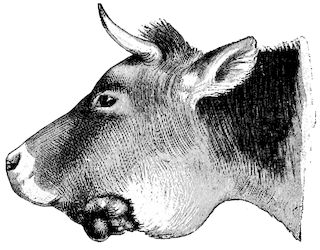
Fig. 268.—Actinomycosis of the jaw.
The earliest symptoms consist in swelling of the bone, which may be overlooked if within the mouth, but the outline of the jaw soon becomes deformed, generally in the middle region of the row of molars. Somewhat tender and firm to the touch at first, the tumour gradually increases in size, invades the deeper regions of the skin, and displays fluctuation at one or two points, followed by abscess formation. The pus discharged may be white, creamy, and inoffensive, but the cavity of the abscess shows no tendency to cicatrise, and the opening through which the pus has escaped is transformed into a fistula. From this moment the pus discharge is of a greyish, sanious nature, and contains a greater or less number of little yellowish grains. It soon acquires an offensive odour, and the fistulous opening is surrounded by exuberant granulations, forming a fungoid mass.
The neighbouring tissues become hardened and lose their sensitiveness, the jaw becomes completely deformed, and a condition is set up which the old writers considered as true cancer of the jaw or maxillary osteosarcoma (Fig. 268).
A probe passed into the fistula penetrates deeply, usually into the thickness of the jaw itself, and however carefully manipulated injures the diseased tissues and causes free bleeding.
If neglected, these lesions become steadily worse, mastication is more difficult, being possible only on the healthy side, and the animals lose condition and eventually die of exhaustion. The external lesion, represented by the fungoid mass, increases in size, assumes a blackish colour, and discharges an offensive liquid. Portions of it undergo mortification and give off a characteristic and extremely fœtid odour. The molars become loose and in some cases fall out, but development is 674usually slow, and some weeks or months elapse before this stage is reached.
When the disease attacks the region of the incisors the symptoms are much sooner apparent, and treatment is much easier. The parasitic invasion results from an injury to the jaw caused by shedding of the milk teeth. Swelling of the body of the jaw thrusts the lower lip downwards, interferes with the prehension of food, and calls for prompt treatment. The disease is rarely allowed to attain the degree of development shown in Figs. 269 and 270.
As in the preceding instance, the animals die of exhaustion unless relieved.
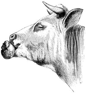
Fig. 269.—Actinomycosis in the region of the incisors.
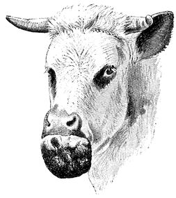
Fig. 270.—Actinomycosis in the region of the incisors.
For reasons difficult to explain, but probably because inoculation is less easy, actinomycosis is much rarer in the upper than in the lower jaw. The disease develops exactly as above described, but shows much less tendency to external ulceration. It invades the maxillary sinus and the region of the palate, and fistulæ are found opening into the buccal cavity, while at the same time the region of the forehead is often deformed.
ACTINOMYCOSIS OF THE TONGUE.
Actinomycosis attacks the tongue apart from any lesion of the jaws, and produces what is commonly called “wooden tongue.”
The disease develops in the tongue itself, generally in the submucous zone, and causes chronic interstitial inflammation, infiltration 675of the connective tissue, and, in time, changes in the muscular structures themselves.
The tongue shows progressive hypertrophy, and becomes hard, sensitive, rigid, and incapable of free movement. As a result the patients first have difficulty in grasping food, then in swallowing their saliva, which dribbles from the mouth, and finally are quite unable to feed themselves.
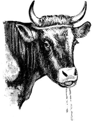
Fig. 271.—Actinomycosis of the tongue.
The tongue is enlarged and indurated, and fills the entire cavity of the mouth. Sometimes it projects beyond the incisors, excoriated and bleeding. On passing the hand into the mouth it is found that the surface is covered with little yellowish or red ulcerated nodules, varying in size from that of a large pin’s head to that of a lentil.
In eating, the animals seize food between the lips and lift the head high, so as to allow the food to fall between the rows of molars. The motion is very similar to that of a fowl drinking.
ACTINOMYCOSIS OF THE PHARYNX, PAROTID GLANDS AND NECK.
Actinomycosis may sometimes leave the mouth and tongue unaffected and attack the pharynx, from which it extends in the direction of the parotid glands and external surface of the neck. In these cases, however, the inoculations are more localised than when the surface of the tongue is attacked, and the lesions consist of vegetations, polypi, or actinomycomata.
The growths develop on the posterior pillars of the fauces, on the sides of the pharynx, or near the entrance to the œsophagus. They interfere with swallowing, and produce symptoms which are easy to detect and interpret.
The lesions may also affect deeper seated tissues and produce growths in the parotid or subparotid region, or lead to the development of fistulæ in the region of the neck. Most fistulæ, however, in 676this region are due to specific inoculation of external injuries. Fistulæ originating in the parotid region and in the upper part of the neck usually resemble in appearance the maxillary fistulæ. The external fungoid growth, however, is less exuberant, suppuration is less abundant, and the surrounding induration less extensive.
Various localisations. Although the disease generally attacks the mouth, tongue or pharynx, it may invade the œsophagus, rumen, reticulum, liver and intestine, larynx, trachea, lung, peritoneum, epiploon, and even the udder.
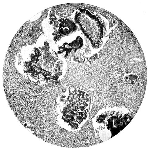
Fig. 272.—Actinomycosis of mammary gland (cow).
Localisations in the udder and peritoneum are commonest in pigs, and it is believed that inoculation occurs either through the galactophorous sinuses or through the abdominal wounds made for purposes of castration.
Causation. The cause of actinomycosis is to be sought in the development of Actinomyces bovis within living tissues. It seems problematical whether the germs to be found in the pus or saliva of affected animals ever directly infect new hosts, and it is difficult to carry out infection in this manner even in very sensitive experimental 677animals. Nevertheless, the persistence of the disease in certain byres would seem to support the view of direct infection.
On the other hand, it is proved that the actinomyces is a parasite affecting vegetables, principally the graminaceæ, and that domestic animals are most commonly infected through injuries caused by vegetable substances. This is suggested by the discovery of the débris of grain at the point where the lesions have originated.
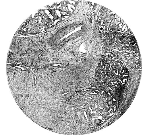
Fig. 273.—Actinomycosis of mammary gland (cow), showing growth invading lobules. (Figs. 272 and 273 are from blocks kindly supplied by Mr. Gilruth, F.R.C.V.S.).
Inoculation is commonest in the mouth and on the surface of the tongue, parts which are, so to speak, permanently excoriated. The shedding of the temporary molars favours such accidents, and this is why actinomycosis of the jaw is, relatively, so common. The incisor region may also be inoculated during the shedding of the milk teeth, but as the infected food comes more closely and for much longer periods in contact with the molars, it is easy to understand why actinomycosis is rarer in the incisor region.
The conditions are less favourable for inoculation of the pharynx, because food does not remain in position there for more than a 678second or two, but when the epithelium has been shed as a consequence of laryngitis or pharyngitis, infection may occur.
As regards cutaneous inoculation, the parasite only seems dangerous when the skin is excoriated or injured either accidentally or as the result of surgical interference.
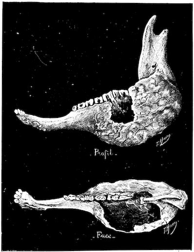
Fig. 274.—Old-standing bone lesions in a case of actinomycosis of the jaw.
Actinomycosis of the lung is probably caused by the germs being inhaled along with the inspired air.
Lesions. The lesions are very peculiar in character, and end in completely destroying the tissues invaded.
Once lodged within an organ, the disease shows a tendency to extend in all directions, and, despite the defensive reaction of the tissues, it soon forms numerous parasitic centres.
In bones, for example, actinomycosis invades the spongy tissue with the greatest ease. It causes subacute ostitis, which leads to diffuse suppuration and local hypertrophy of the bone, destruction of the compact layers, and the development of an abscess with fungoid, exuberant, granulating walls which show no reparative tendency whatever.
The pus of the abscess and the liquid from the fistula contain varying quantities of yellowish grains, representing clusters of actinomyces. The surrounding tissues, muscles, tendons, skin, etc., are all involved before long in the inflammatory process, and the granulating masses themselves are invaded by the yellowish parasitic tufts. All the fistulæ are surrounded by enormous zones of infiltration, which on 679incision exhibit a lardaceous appearance. On section it may appear that the lesion is confined entirely to the bone, though this is exceptional (Fig. 274). Ordinarily the neighbouring tissues are also destroyed, and not infrequently there is communication with the external air. Sections then display a fungoid tissue, interspersed with perforated lamellæ of bone and lardaceous tissue containing cavities crammed with actinomyces.
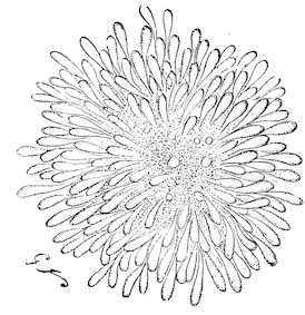
Fig. 275.—Highly-magnified clump of actinomyces.
The lesions in the parotid regions, the neck or other parts attacked always present the same appearance, viz., wide, tortuous, bifurcated fistulæ, with exuberant granulations both in the direction of the cavities and of the exterior, together with lardaceous induration of the tissues and abundant fœtid liquid pus.
When it affects the tongue the parasite is to be found in the submucous region, where it causes little swellings, which, when superficial, rapidly undergo ulceration. The subjacent regions, the interstitial connective tissue, and the muscular tissue become infiltrated, hardened and progressively sclerosed. The tongue is gradually hypertrophied, and soon it becomes as hard as wood, whence the term “wooden tongue.”
Actinomycosis of the lung may easily be mistaken for tuberculosis, for the centres, although usually confined to one lobe, may also be disseminated. The lesions, however, are surrounded by an abundant fibro-sclerous inflammatory tissue.
In the abdominal cavity, particularly in sows, actinomycotic lesions occur as little masses varying in size between that of a pea and that of a haricot bean, attached to the epiploon and peritoneum and filled with pus containing mycosic grains.
Diagnosis. Actinomycosis is usually easy to recognise, both on account of the special character of the lesions and the presence of the little grains formed by the parasite. The practitioner will rarely fail to recognise at once the signs of actinomycosis of the jaw, but actinomycosis of the tongue is more apt to be mistaken for deep-seated sclerosing glossitis, although a careful examination will always enable the different symptoms to be distinguished.
680It is otherwise with regard to growths in the pharynx and œsophagus, for, until after removal, simple polypi cannot be distinguished from actinomycotic growths. In such cases the administration of iodide of potassium affords valuable indications.
The prognosis is grave, whatever the clinical form of the disease. Important advances, it is true, have lately been made, and the iodide of potassium treatment is of great value, but too much must not be expected of it, and its benefits have certainly been exaggerated. Clinical experience suffices to prove that only actinomycosis of soft tissues can be cured by drugs, bony lesions being amenable only to medical and surgical treatment combined. Even combined treatment is often unsuccessful.
Treatment. Thomassen in 1885 first explained the favourable action of iodide of potassium on actinomycotic growths, and Nocard in 1892 again directed public attention to the advantages attending the use of this drug both in man and the lower animals. As too frequently happens, however, the benefits of this treatment have been exaggerated, and iodide of potassium has been held out as a specific even against lesions in bone. With very few exceptions this is incorrect, and, as Moussu has shown, when the disease affects bone tissue it only yields to mixed treatment.
The treatment of actinomycosis may therefore be considered under two heads.
Firstly, the treatment of actinomycosis of soft tissues; and, secondly, that of bone.
Actinomycosis of soft tissues, muscle, skin, lymphatics, serous membranes, etc., comprises the most common forms of actinomycosis, viz., those of the tongue, pharynx, parotid glands, neck, etc.
The second form comprises actinomycosis of the lower jaw (molar region), the upper jaw, region of the incisors, etc.
Iodide of potassium in daily doses of from 2 to 3 drachms is almost a specific in dealing with the first form of disease.
In lingual actinomycosis, for example, the effects may be seen a few days after treatment is begun. The tongue becomes softer and more mobile, can be protruded beyond the mouth and retracted into it, and day by day tends progressively to resume its normal appearance.
The patients, which were previously slowly dying of inanition because they were unable to feed themselves, again take to their food and begin to put on flesh. To ensure the treatment being efficacious it should, as a rule, be continued for three or four weeks.
During the course of this treatment the system becomes saturated with the drug, but no bad effects follow. The patients suffer from lachrymation, coryza, bronchorrhæa, and especially iodic eczema, but 681all these symptoms diminish and disappear soon after the administration of the drug is discontinued.
Recovery, however, is not always permanent, and even when the tongue has resumed its normal appearance a relapse may occur. We have seen several such cases after treatment extending over more than six weeks, and it is therefore often advisable to fatten the animals as rapidly as possible and prepare them for slaughter.
If no relapse occurs, and recovery is regarded as permanent, another complication may make its appearance, viz., sclerous atrophy of the tongue. This is almost as dangerous as the primary lesion, because it prevents the animals from feeding, and constitutes an additional reason for following the course above suggested.
Other lesions of soft tissues, such as disease of the parotid or cervical glands, etc., yield to the same treatment, but it is advisable first of all to clean out the fistulæ, scrape off exuberant granulations, cleanse the irregular culs-de-sac, and thoroughly curette all accessible parts.
Treatment is much longer than in the case of actinomycosis of the tongue, but it is not always necessary to push the remedy to extreme limits. As soon as symptoms of iodism appear only a drachm or two of the drug need be given daily.
Actinomycosis of Bone.—As a general rule, actinomycosis of bone resists the administration of iodide of potassium, a fact probably explained by the much less abundant blood supply in bone as compared with very vascular tissues, such as the tongue.
To have any chance of success the iodide treatment must be supplemented by surgical interference. As regards the surgical aspect of the case, the affected bone should be removed as far as possible, together with all broken down tissue. Should this be neglected, the disease returns in a little while.
In actinomycosis of the region of the incisors the method is radical when adopted in time. The body of the maxilla can be partially removed with a fine saw, two cuts being made disposed thus: < (the letter V sideways). The upper and lower layers of compact tissue should be spared as much as possible, so that the body of the bone may not afterwards break. Recovery is only a matter of time.
A very small local iodoform dressing is applied, and, when healthy granulations appear, cicatrisation can be left to natural means.
Cases of actinomycosis of the jaw are much more troublesome. If, as usually happens, the lesion is ulcerated before the practitioner is called in, the external fungoid growth should be removed by means of an elliptical incision through the skin, the axis of the ellipse being parallel with the branch of the maxilla. The bony fistula is then exposed.
682In following up this fistula care must be taken not to injure the facial artery, the facial vein, or Stenon’s duct. Once the bone is exposed the disease can be attacked in the depths. The diseased interior is cut away by means of a special curette, all affected portions being removed, and an iodine or iodoform dressing is then applied.
The operation is extremely troublesome, owing to the enormous bleeding, and sometimes it is impossible to carry out successfully, as in the case of old-standing and extensive lesions. To ensure recovery under such circumstances, it is necessary to remove a portion of the branch of the jaw, and this, though quite possible from the scientific standpoint, would not be worth while in an animal, the value of which is usually small.
Curettage of the bone is only of value in dealing with recent lesions, and even then should not be practised except in the case of animals which the owners particularly desire to keep.
In cases of actinomycosis of the upper jaw surgical treatment is just as difficult as in the lower jaw, and calls for similar precautions.
The diseased portions of bone having been removed, the cavity is plugged with iodoform or cotton wool, or a dressing saturated with boric acid and iodoform.
In all surgical operations it is important not to injure the dental arteries or nerves, or the alveolo-dental periosteum.
TUBERCULOSIS.
Tuberculosis is a contagious disease produced by the action of Koch’s bacillus. It is common to man and all domesticated animals, but it specially affects animals of the bovine species. Its existence has long been recognised, although in oxen it was formerly confounded with the lesions of peripneumonia and echinococcosis.
It was not until the beginning of the nineteenth century that Laënnec (1811) described the tuberculous lesion from the anatomical and pathological standpoint. Gürlt pointed out for the first time in 1831 the similarity, the identity in fact, of tuberculous lesions in man and the ox.
In 1865 Villemin showed that tuberculosis could be conveyed from animal to animal, always producing similar lesions, and in 1868 Chauveau proved that, in the calf, infection might arise simply from the eating of tuberculous material.
At a somewhat later date doubts were entertained regarding the identity of human and bovine tuberculosis. Virchow denied the identity of the two diseases on the basis of a comparative study of the lesions. His opinion, however, has not prevailed, and the doctrine 683of the identity of tuberculosis in mammals still appears probable, in spite of the recent declarations of Koch (1901).
Causation. Tuberculosis is due solely to the activity of the tubercle bacillus. In 1884 Koch isolated and cultivated this bacillus in living animals, and always reproduced typical tuberculous lesions by injecting cultures. In 1887 Nocard and Roux described a rapid method of cultivating the bacillus, and in 1890 Koch announced the discovery of tuberculin.
The tubercle bacillus assumes the form of a little rod, five or six micromillimètres in length, and ·03 to ·05µ in thickness. It has a special staining reaction when treated with Ehrlich’s or Ziehl’s solution. It grows between 98° and 104° Fahr. (37° and 40° C.) in various artificial media containing glycerine.
Healthy subjects become infected by the accidental entrance of germs into their bodies, either by the respiratory and digestive tracts, or through solutions of continuity in the skin.
The material from tuberculous centres is virulent, whether consisting of sputum or discharge, saliva, fæces, urine, milk, etc., or tuberculous tissues derived from the different viscera.
The blood and muscular tissues are not always virulent, even in cases of generalised tuberculosis.
The virulent organisms usually enter the body through the lymphatic system; invasion proceeds from the point inoculated towards the nearest lymphatic glands and thence along the chain of lymphatic vessels, and the lesions extend, attacking the internal organs more or less rapidly. The body does not necessarily become fatally infected as a consequence of accidental or even experimental infection, for the bacillus may itself be destroyed by the phagocytes, or the lesion may remain purely local.
Although tuberculosis is the gravest and most widespread disease on the surface of the globe, its contagious character is relatively little marked, a fact which has unfortunately led to its receiving little attention in ordinary life.
Contagion is usually the result of cohabitation, although contact between diseased and healthy subjects for a period of some days or even weeks does not seem sufficient to produce the disease. Nocard has fixed a mean period of five to six months as necessary for the contraction of the disease by bovine animals, and Moussu has arrived at almost identical results by placing tuberculous and healthy cows together in a byre reserved for such researches. In this connection, however, very great differences of individual susceptibility exist, and these are difficult to appreciate in the present state of our knowledge. It thus happens that an animal of vigorous appearance and in good 684condition may easily contract tuberculosis, whilst a thinner and less vigorous one will resist it for a comparatively long time.
Speaking generally, it may be said that young animals contract tuberculosis by cohabitation in infected places more easily than adult or aged ones, and the fact that old animals contribute the larger number of cases is to some extent due to their having in the course of their lives been more exposed to continued or successive infection.
Contagion does not occur in byres unless as the result of the presence of animals with open tuberculous lesions, such as caverns in the lungs, tuberculous bronchitis with ulceration of the mucous membrane, tuberculous metritis, enteritis, etc. The virulent germs are expelled in the saliva, nasal discharge, excrement, etc., and are distributed over the forage, manure, litter, and in the drinking water; after desiccation they may be spread by currents of air.
The mangers, racks, drinking pails, and various stable utensils become permanently contaminated, the air of the cowsheds contains virulent dust, and the animals there confined are continually exposed to infection either through the respiratory or digestive passages.
Contamination through the respiratory tract is by far the most frequent cause of the evil, and recent experiments at Pouilly-le-Fort (1900) have shown how easy it is to convey the disease experimentally by inhalation.
Patients suffering from closed tuberculous lesions of the pleura, pericardium, spleen, peritoneum, etc., do not spread the bacilli. Healthy animals may remain in contact with them without danger, but it is well to remember that such cases are quite exceptional. As a rule the lesions are of a mixed character, and the general principle may be laid down that cohabitation of any duration with tuberculous subjects is dangerous.
Contagion spreads more easily, in proportion to the number of tuberculous subjects in a given byre, to the total number of animals in a herd, and to the neglect of cleanliness, good feeding, ventilation, etc.
Life in the open air and at grass greatly diminishes the chances of contagion. The virulent products are then disseminated in all directions and are soon destroyed by the general atmospheric conditions. Close confinement in ill-ventilated stables, on the contrary, strongly tends to the propagation and development of tuberculosis.
In calves infection may occur through the alimentary tract by means of tuberculous milk, whether such milk is obtained directly from the udder or out of a pail. The same may be true of young pigs fed with skimmed milk.
Goats contract tuberculosis somewhat readily by confinement in 685byres with tuberculous cows, and Moussu declares that contagion afterwards spreads just as rapidly among goats as among cows. The vaunted great resistance of goats to tuberculosis, formerly so often spoken of, and by some wrongly considered as a condition of immunity, is deceptive, and if tuberculosis is less frequently seen in goats, this is solely because goats enjoy the greatest liberty at all seasons.
On the other hand, the disease is very rarely conveyed to sheep, even when they are kept for long periods with tuberculous cows. Moussu found that two years of close cohabitation were necessary for its development under these conditions.
Heredity is a factor of the highest importance in determining the causation of tuberculosis. At the present time a tendency exists to deny this, but such a view is erroneous.
Observation has clearly shown that tuberculosis is rarely conveyed from the mother to the fœtus, and that practically none of the calves borne by tuberculous mothers react to tuberculin (95 per cent.: Nocard and Bang); but even if this is absolutely correct, it only shows that great benefits might be derived if proper sanitary organisation and intelligent hygienic conditions in byres were found everywhere in the country. Unfortunately in practice this is far from being the case. These non-tuberculous calves are left in common contaminated byres, where they rapidly become infected and perpetuate the disease.
Physiologically these facts are easily explained. The placenta resists the passage of microbes, or at least only allows them to pass under quite exceptional conditions, and practically only when the blood-vessels are affected. As, on the other hand, tuberculosis of the ovaries, Fallopian tubes or uterus generally prevents pregnancy and causes sterility, there is nothing extraordinary in the fact that tuberculosis is not hereditary in the strict sense of the term. The influence of the sire has been invoked, but it has been proved that direct paternal infection is only possible where ulcerating tuberculous lesions of the testicle, prostate, or vesiculæ seminales exist. Such conditions seldom or never occur in the sires of domestic animals.
As a general rule, therefore, it may be said that tuberculosis is not hereditary. New-born animals become infected during the months following birth, either directly through the alimentary tract when the mothers are suffering from mammary tuberculosis, or, perhaps more frequently, through the respiratory and digestive tracts.
But although microbic infection is not hereditary, it by no means follows that the offspring of tuberculous subjects are as well prepared for the struggle of life as the descendants of healthy subjects. What is transmitted is a greater tendency to contract the disease.
686This aptitude or predisposition is of such importance that in Moussu’s opinion it should be regarded as one of the essential factors in the development of tuberculosis. The cause of tuberculosis is Koch’s bacillus. It does not always produce its full effects in animals born of healthy parents; but in one that suffers from a tuberculous hereditary taint tuberculosis appears.
Physiological and pathological researches cast considerable light on this question. In tuberculous mothers the organism not only suffers from the infection, but from a permanent intoxication which interferes with normal metabolism in the vital organs and the exchanges between mother and fœtus. If the microbes remain confined to the system of the mother, their poisons are conveyed by the blood and pass through the placental barrier. In a greater or less degree they saturate the tissues of the little creature in process of development, and communicate to it a peculiar hereditary taint. The effects of this taint are often noticeable from the moment of birth, for comparative physiological and pathological investigations have shown that the tissues of tuberculous animals assimilate given foods less perfectly and are the seat of greater losses of all kinds than those of healthy subjects.
Although the disease itself, therefore, is not hereditary, it is otherwise with the organic taint which plays so important a part in its development. This organic taint consists in a special condition of the tissues or cells of the parents, which show a diminished power of resistance to the action of the germs of tuberculosis; it is therefore easy to understand how important a part these influences may play under certain conditions.
Without doubt, in the case of bovine animals, the predisposition could be neutralised in carefully managed studs by the immediate isolation of the new-born under conditions which shield them from tuberculous infection, and experiment has shown the benefits derived from such precautions; but it must not be forgotten that intelligently managed studs are the exception, and that for a long time to come we must in practice take cognisance of the actual conditions under which the disease develops.
The lesions of tuberculosis vary greatly in appearance, according to the organs affected, though the method of development is always identical.
The primary lesion corresponds to what has been termed tuberculous granulation, or anatomical tubercle properly so called; this, the macroscopical, pathological entity, assumes the form of a small prominent centre, semi-transparent, greyish, opaque or yellowish, according to its age.
687These tubercles, produced by the presence of colonies of bacilli, are due to the defensive reaction of the invaded tissues, which gradually undergo change and are destroyed in a direction radiating from the centre towards the periphery. The tubercle in itself has no very specific character—only the bacillus.
The elementary lesion may remain isolated, but very frequently it is closely surrounded by other similar tubercles, and becomes enveloped in a common inflammatory area. A large portion of an organ may appear as if riddled with tubercles of different age and size, while the interstitial connective tissue reacts and forms fibrous separating partitions. The general appearance is that described under the term “diffuse tuberculous infiltration.”
At a still more advanced stage in the development of the disease conglomerations are produced, consisting of tuberculous masses the size of a hazel-nut, a walnut, an egg, a man’s fist, or even larger. These lesions, irrespective of size, undergo caseous degeneration from the centre towards the periphery.
In exceptional cases the tubercles remain fibrous. More frequently, particularly in animals of the bovine species, they become infiltrated with lime salts. Caseous degeneration not only invades the centre of the tubercles but also the peripheral layers, and sometimes the whole of a conglomerated mass.
Steadily pursuing their course of pathological development, the tuberculous masses become softened and are transformed into tuberculous abscesses, which open towards any free passage, leaving behind sometimes ulcerations, sometimes caverns of varying sizes, or blind simple or bifurcated fistulæ.
Recent experiments by Nocard and Rossignol (1900) prove conclusively that a certain time (always more than a fortnight) elapses between the moment of entry of the contagion into the organism and that at which its effects become manifest by furnishing a reaction to tuberculin. Calcification or softening of the lesions, moreover, never occurs in less than fifty days.
According to the organs studied, these tuberculous lesions assume certain appearances, which in each locality seem almost always to be identical.
Thus, as regards the larynx, trachea, and bronchi, the tubercles develop in the depths of the mucous membrane, rapidly undergoing caseous transformation, softening and purulent degeneration, and producing numerous isolated or confluent ulcerations in the air passages.
According to the case and the kind of animal affected, the lung presents either disseminated tuberculous formation, tuberculous infiltration, tuberculous conglomeration, or cavern formation.
688The lung may be affected to such a degree that it appears incredible that the blood can have been sufficiently aerated to support life.
The lungs may be transformed into yellowish, caseous, calcareous, or softened masses enveloped in thick, fibrous, resistant walls. The intervening pulmonary tissue may be healthy in appearance, or reddened, congested, and sometimes hepatised.
The pleural, pericardial, and peritoneal membranes may be covered with exuberant tuberculous lesions, like ripe mulberries, in consequence of fusion and massing of the tuberculous growths. The primary tubercles are surrounded with fibrous walls, which granulate when on the surface of a serous membrane, and impart to the membrane a vegetative, sometimes villous appearance, and a colour varying from pink to light or dark red.
The collective lesions lining the cavities are described by butchers under the significant term of “grapes.” In the interior of these exuberant masses, which sometimes form layers an inch or more in thickness, the tuberculous lesions undergo the usual developmental changes, that is to say, they become caseated or infiltrated with lime salts, but they do not so readily undergo softening as those of the lung. The parietal and visceral serous membranes readily become adherent at numerous points, setting up union between the lung and the walls of the chest, or the intestine and the walls of the abdomen, etc.
In the pericardium the vegetations are frequently of a fungoid character.
Tuberculosis of lymphatic glands sometimes assumes a disseminated, discrete form or that of a diffuse infiltration, or, again, in old-standing cases it constitutes a massive tuberculous conglomeration. In point of fact, the lymphatic glands as such no longer exist, their tissue having undergone total degeneration; they are represented only by an enlarged, thick, fibrous shell, forming the envelope which encloses caseated and calcareous masses of a more or less soft nature.
Tuberculous infiltration of the submaxillary and sub-parotideal lymphatic glands interferes with swallowing and breathing, compresses the pharynx, œsophagus and larynx, and deforms the head.
Compression of the arteries, veins, nerves, etc., at the entrance to the chest may cause various symptoms which are not difficult to interpret. The glands at the entrance to the chest and the whole of the anterior mediastinum may form a single mass. Lesions in the posterior mediastinum, however, are of even greater importance and explain certain symptoms, such as difficulty in swallowing, spasm of the œsophagus, mechanical contraction of the œsophagus, permanent tympanites, etc., for which the state of the lungs alone would not account.
689Even when the lungs are unaffected it may happen that the lymphatic glands of the mediastinum (superior or inferior œsophageal lymphatic glands) and the bronchial lymphatic glands may be so diseased that the œsophagus is completely surrounded and compressed by them, and its function thus seriously impaired (Fig. 276).
In the abdomen the mesenteric glands are most exposed to disease, and when infected through the intestinal tract they assume the form of large flattened masses arranged along the mesentery.
In the digestive tract, as in the trachea and bronchi, tuberculosis has a marked tendency to assume the ulcerative form. Disseminated or aggregated tubercles develop in the thickness of the mucous membrane, and, after rapidly softening, become ulcerated. The nature of these lesions can only be determined by noting their character and examining the discharge.
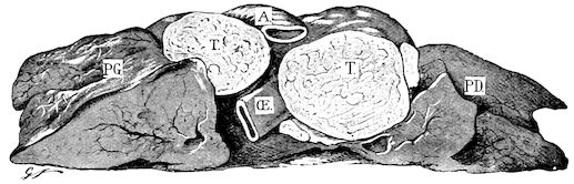
Fig. 276.—Tuberculosis of lymphatics. PG, Left lung; PD, right lung; TT, tuberculous œsophageal lymph glands; A, aorta; Œ, œsophagus (the lung is divided transversely near its centre).
The ulcerations are localised in the mouth and pharynx, in the second half of the small intestine towards the ileum, and in Peyer’s patches.
Tuberculous lesions develop in the vaginal sheath of the male genital organs exactly in the same way as in an ordinary closed serous cavity; tubercles may also develop on the surface or in the substance of the testicle. They become aggregated, undergo softening, spread towards the interior, and may break down, thus forming abscesses. In the female genital passages the disease invades the thickness of the walls, but shows a marked tendency to ulceration, as in the intestine or trachea.
In the udder tuberculosis is generally diffuse, shows a tendency to hypertrophy and the free formation of fibrous or sclerous tissue; only tubercles in the glandular layer of the acini become ulcerated. In time the whole of the secreting structure undergoes diffuse tuberculous suppuration, fibro-caseous masses form in the depths of the tissue and may soften, producing deep-seated tuberculous “cold abscesses.” 690The mammary lymphatic glands are affected in the same way as other lymphatic glands.
In the joints tubercles appear either on the synovial membrane or in the thickness of the bony epiphyses, very often at both points simultaneously. The synovial membrane is covered with vegetations and villous growths, the ends of the bones are attacked by a destructive ostitis, tubercles or tuberculous centres form in the thickness of the spongy tissue, the articular cartilages are destroyed, the ends of the bones become deformed, and in the last stages fungoid arthritis in various forms may be produced.
In bones the tubercles originate in the depths of the spongy tissue. They produce destructive hypertrophic ostitis, in which the bony tissue is replaced by tuberculous centres or masses divided by fibrous partitions. On section, these lesions exhibit the same yellowish caseated or calcified appearance as the lesions of other affected organs. The compact layer may sometimes be perforated at several points before being destroyed.
In tuberculosis of the brain the primary lesions develop at the expense of the serous layers of the arachnoid and on the pia mater, towards the base of the brain and the fissure of Sylvius, or at the expense of the small vessels which penetrate the depths of the nerve substance itself. Some tubercles remain isolated, become confluent or are collected in masses of different sizes, and provoke symptoms which vary with the locality attacked.
Symptoms. Tuberculosis is the most protean of all diseases, and at first sight it often seems impossible to assign to one group, clinical conditions presenting such essentially different appearances. All the tissues may be attacked, from the bones to the most delicate of the viscera, a fact which explains why all aspects of tuberculosis cannot be described. Certain forms, however, occur very frequently, and may be regarded as classic; these will be considered in the order of their frequency.
TUBERCULOSIS OF THE RESPIRATORY APPARATUS.
Without doubt this form of tuberculosis is by far the most frequent. It assumes the form either of bronchitis, laryngo-bronchitis, or pulmonary tuberculosis.
Tuberculous Bronchitis.—The symptoms of tuberculous bronchitis do not essentially differ from those of ordinary bronchitis, though the disease develops more insidiously and slowly, and is seldom accompanied by fever. At first the cough is dry and suppressed; later it becomes paroxysmal, and at a still more advanced period liquid and 691rough. The least irritation brings on these attacks of coughing; changes from the warmth of the stable to the coldness of the outer air or vice versâ, the presence of dust or the action of liquids when drinking, etc., etc. During the first stage coughing is not followed by expectoration, but later yellowish-grey, glairy mucus may be discharged: more frequently it is coughed into the pharynx and swallowed.
These symptoms continue for weeks or months without showing any tendency to abate. If the larynx is attacked inspiration becomes rattling and difficult, while the neck and head are held extended, and the least pressure over the larynx produces coughing.
Tuberculosis of the larynx, trachea, and bronchi is usually accompanied by disease of the lung, but may occur by itself.
When there is a discharge it consists of thick, viscous, sticky mucus of a peculiar greyish-yellow colour. Microscopical examination shows it to contain tuberculous bacilli.
Pulmonary Tuberculosis usually assumes the chronic form, and is almost always preceded by specific bronchitis. The patients retain their appearance and condition for a longer or shorter time, and, without the experience resulting from continued observation, it would be difficult to believe them to be suffering from the slow development of a serious disease.
Frequent coughing without any apparent reason is the only symptom likely to arouse suspicion.
At a later stage these animals lose condition, feed less eagerly or exhibit capricious appetite, and sometimes well-marked and repeated digestive disturbance, such as slight tympanites with constipation or diarrhœa, moderate impaction of the rumen, relative atony and slackening of peristaltic movements. The wasting gradually becomes more marked or, in the case of pregnant or milch cows, makes intermittent progress, until the animals become anæmic and finally cachectic. The cough is more frequent and more severe, and is followed by discharge from the nose or by swallowing movements. From this time phthisis, properly so called, exists.
The course of the disease is not invariable. Certain animals may appear ill for years without clinically showing the least apparent aggravation; others on the contrary, though living under similar conditions, are rapidly attacked, and in six to twelve months exhibit all the signs of advanced phthisis. Pregnancy, suckling, and prolonged lactation favour the development of the disease by taxing the physical resources of the animal.
Animals suffering from phthisis exhibit a peculiar appearance. They are extremely thin, all their soft tissues are wasted, the limbs are dragged in moving, respiration is rapid and sometimes jerky, 692the mucous membranes are pale and discoloured, and the skin is tight and adherent to the subjacent tissues.
These general signs, however, would not warrant a diagnosis, for, apart from the cough, certain other diseases present all the external appearances of the last period of tuberculosis (chronic diarrhœa, chronic forms of poisoning—bacterial or otherwise—dyspepsia, etc.).
In cases of doubt it is essential to discover by percussion and auscultation that the external signs are really the result of lesions of the lung, and that the lung disease has developed gradually in accordance with the signs shown by simple external inspection.
The symptoms presented during the development of the pulmonary lesions may be divided into three phases.
In the first phase percussion gives no information, though auscultation reveals rough respiration, inspiration and expiration being also unequal. Expiration, which, in the healthy subject, is silent, becomes clearly perceptible, not over the whole lung, but usually over the anterior lobes, particularly the cardiac lobes. This sign is the result of tuberculous infiltration and of the neighbouring pulmonary tissue having lost its elasticity.
Inspiration is rough and rasping, and sometimes occurs in several stages, the act being interrupted or jerky; expiration lasts longer than inspiration, is rough and prolonged, but never blowing in character. These peculiarities are only found in one other condition of the lung, viz., emphysema.
The patients appear little affected in this, the first, stage of tuberculosis. But for the cough they may seem perfectly healthy.
In the second phase the tuberculous infiltration extends and ends in the massing, by fusion or centrifugal growth, of the tuberculous masses.
Percussion may now indicate localised dulness, but this is not invariable, because the diseased anterior and middle lobes of the lung are concealed beneath the muscles of the shoulder. When dulness is noted, it is usually over the lower part of the posterior lobes, very rarely at any higher point on the side of the chest. Frequently the dulness is only partial.
On auscultation the signs met with during the first stage become much more marked. Inspiration is always rough, rasping, painful and difficult at certain points, particularly in the anterior zones. In this region expiration is rough, prolonged and sometimes of a clearly marked blowing character. This is particularly the case in the subscapular zone and the auscultation zones 2 and 3 (Fig. 166). In the dorsal region and in zone No. 1, respiration may appear normal. Nevertheless, the sounds are propagated to a distance, the 693infiltrated lung steadily loses its elastic qualities, the vesicular murmur entirely disappears from the affected regions, and the sounds noted are of bronchial origin.
Like the first, the second phase may vary in intensity, extent, and in the diffusion or localisation of the tuberculous lesions. Blowing respiration may be noted over different areas, accompanied by sibilant, snoring and migratory mucous râles. The vesicular murmur is exaggerated in the healthy parts, coughing, accompanied by expectoration or followed by swallowing movements, is frequent, the appetite becomes capricious, and the general condition suffers. In this second phase almost the whole of one lung may be diseased and exhibit the signs described.
The third phase corresponds to the softening of the tuberculous masses, and the formation of ulcers and caverns. The zones of dulness or partial dulness may be more extensive, though cavern formation is usually confined to the anterior or middle lobes. Percussion still affords no precise information.
As the tuberculous masses undergo softening and ulceration, their contents are gradually passed into the bronchi, and auscultation reveals signs indicative of the existence of caverns, which signs vary with the dimensions of the caverns themselves. On auscultation the respiration is always found to have at certain points a blowing character, and it may even develop into a true tubal souffle. In other areas, where the caverns are merely in course of formation, gurgling sounds are all that are heard, but where true caverns exist there is an incessant cavernous souffle.
The lesions peculiar to the third phase are seldom seen in practice; because the animals become anæmic, exhausted and cachectic, they are usually slaughtered early. Nevertheless, the third stage occasionally develops in an astonishingly short time, six to eight months at most.
Very frequently the patients, although cachectic and even phthisical, do not yield on auscultation the sounds described as peculiar to the third stage, because the tendency to softening is not very marked in bovine animals. The lungs exhibit massive infiltration, and, whilst pulmonary consumption is not uncommon, the development of caverns is comparatively rare.
The expectoration or discharge in this third form is puriform, glairy, viscous, and of a dirty-yellow or even greenish-yellow colour. Bacteriological examination reveals the presence of tubercle bacilli and adventitious organisms.
These conditions are always associated with various complications, and the second and third stages of chronic tuberculosis are frequently 694accompanied by lesions of the pleura, of the mediastinal lymphatic glands, of the liver, etc.
Digestive disturbances often occur; the appetite is capricious or in abeyance, there is atony of the rumen and chronic dyspeptic tympanites. These disturbances are easily understood where there are lesions of the liver, intestine, and mesenteric lymphatic glands, but not when the lung alone appears the seat of the disease. In this condition the patients probably suffer from permanent complex intoxication, due to toxins elaborated by the tubercle bacillus and other microbes which multiply on or in the lesions, and this chronic intoxication reacts on the vital functions (innervation, secretion, digestion and nutrition). Nor are the effects limited to these appearances; the heart’s action is also accelerated, and the temperature rises. During the first and part of the second phase there is comparatively little fever, but afterwards this is continuous or of a peculiar intermittent character. In the morning the patient’s temperature may be normal; in the evening it has risen from 1·5 to as much as 9° Fahr. (1·1 to 5·2° C.) above normal, and this recurs day by day. These attacks coincide with softening of the lesions, and when suppurating caverns exist they are more marked and more nearly continuous, assuming the characters of the hectic fever shown in consumption.
Often during the febrile periods the urine is albuminous.
In chronic tuberculosis of bovine animals bleeding from the lung is rare even when caverns exist, and Moussu, in spite of extensive experience, has seen only two cases. This is in striking contrast with the condition in human sufferers from pulmonary tuberculosis, two-thirds of whom bleed at the lungs.
TUBERCULOSIS OF SEROUS MEMBRANES.
After pulmonary tuberculosis, tuberculosis of the pleural and peritoneal serous membranes is the most frequent clinical form of this disease. Sometimes both forms exist, and although the pleural and peritoneal lesions predominate or alone attract attention, there are also lesions in the lung or mediastinal lymphatic glands.
It is difficult to explain how the pleural and peritoneal serous membranes can be seriously invaded without the lung becoming affected, though in point of fact such a state of things frequently exists.
Tuberculosis of the pleura without pulmonary lesions is suggested by very obscure symptoms. The general signs consist in diminution of appetite, loss of condition, tachycardia, elevation of temperature, 695and progressive organic wasting. These are always present, though in themselves they have no specific significance.
The local symptoms are still more vague. Percussion causes pain, and the practitioner might at first suspect peripneumonia. The patient edges away, and tries to avoid the application of the pleximeter hammer. Firm pressure over the intercostal spaces sometimes causes struggling, and produces indications of abnormal sensitiveness. There is generally extensive partial dulness, sometimes complete dulness towards the lower regions of the chest.
On auscultation the lung may reveal the different indications of chronic pulmonary tuberculosis, or simply diminution of the respiratory murmur at points, accompanied by crepitant, sibilant râles, and moist, crackling sounds. As the anterior portions of the pleural sacs are most commonly invaded, the anterior vena cava is compressed, causing some difficulty in the return circulation, and producing venous pulse, which may extend as high as the parotid gland; there is, however, no swelling of the dewlap.
Respiration is frequent and difficult in consequence of adhesions between the pleura and lungs, which are connected by bands of fibrous tissue of varying extent. Coughing is rarely absent, and if the lung is diseased may be followed by discharge containing numerous bacilli. Otherwise the cough exhibits the pleuritic character, that is, it remains slight, dry, paroxysmal, and painful. The pericardium may be affected as well as the pleura; if the conditions occur simultaneously the venous pulse in the jugulars will be particularly apparent.
The symptoms of tuberculous pericarditis are similar to those of ordinary pericarditis, except that the exudation is less abundant; in a word, the symptoms are those of rather trifling exudative pericarditis.
Tuberculosis of the peritoneum is frequently accompanied by that of the pleura or the abdominal viscera. The lesions are localised on the parietal peritoneum and epiploon, producing in time adhesions between the viscera and walls of the peritoneal cavity, which affect the action of the digestive organs, gradually causing interference with the peristaltic movement both of the rumen and the intestines. The stagnation of alimentary matter favours fermentation, so that the rumen becomes permanently distended. The right flank also is swollen, and the abdomen exhibits a change in shape similar to that in peritonism, which is a constant symptom of tuberculous peritonitis.
As in the thorax, the tuberculous lesions seldom produce extensive liquid exudation, so that ascites does not occur, but on palpation the abdominal walls appear to have entirely lost their pliability and 696to be unyielding and greatly thickened, a point which is the more remarkable as the animals are thinner.
The wall of the abdomen is stiff, incapable of being depressed as in ordinary subjects, and gives to the fingers the sensation of a thick hard covering, through which the subjacent organs and their contents, that is, the rumen, intestine and alimentary material, can no longer be felt. This rigidity is always most marked in the lower abdominal region. The digestive peristaltic movement can no longer be detected, and on auscultation the normal sounds are manifestly much slower than usual.
TUBERCULOSIS OF LYMPHATIC GLANDS.
It might perhaps have seemed more logical to place tuberculosis of the lymphatic glands at the commencement of these clinical divisions of tuberculosis, as when tuberculous lesions, of whatever kind, occur in the lung, pleura, abdomen, etc., the lymphatic glands in the neighbourhood are invariably invaded. In such cases, however, the lesions in question are not the dominant features.
Under this heading must be classed tuberculous lesions which, on the contrary, affect the lymphatic glands in so marked a manner that lesions in other organs may be regarded as secondary. This occurs somewhat frequently, because at the present day there is a tendency to believe that inoculation takes place mainly through the mucous membrane of the pharynx, and thence extends towards the neighbouring lymphatic glands. At any rate, it is unquestionable that tuberculosis of the lymphatic glands may exist quite apart from any other lesion visible to the naked eye.
Two forms are very common, tuberculosis of the retro-pharyngeal region and of the neck, and tuberculosis of the mediastinal lymphatic glands.
Tuberculosis of the Retro-pharyngeal Glands.—In addition to the retro-pharyngeal glands the cervical chain of lymphatic glands, the subglossal, subatloid, preparotid, and even the prescapular lymphatic glands and those at the entrance to the chest, may also be invaded more or less.
This form of tuberculosis may remain latent for a long time, attention being attracted to it only when deglutition is impeded and local deformity becomes apparent.
Swelling of lymphatic glands resulting from tuberculous infection is slow and progressive, differing entirely from that which accompanies suppurative adenitis. The neighbouring connective tissue is certainly somewhat thickened or infiltrated, but the glands themselves can always be detected. The region of the gullet is enlarged, the 697depression marginating the lower jaw is filled up, the subatloid space disappears, the subglossal glands occupy the space beneath the tongue, and in cases where the lesions are very pronounced the œsophagus and larynx may even be pushed downwards.
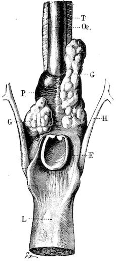
Fig. 277.—Lesions in retro-pharyngeal tuberculosis. T, Trachea; Œ, œsophagus; P, pharynx; H, hyoid bone; E, epiglottis; L, tongue; G, tuberculous retro-pharyngeal glands.
Swallowing is difficult, in consequence of compression of the upper part of the œsophagus, and, as the laryngeal nerves may be included in the swelling, dyspnœa or roaring not uncommonly results.
By palpation with one or both hands it is easy to identify the glands and detect enlargement, hardness and sensitiveness. In exceptional instances the caseous masses they contain undergo softening and conversion into purulent material.
When the cervical lymphatic glands are attacked the jugular furrows disappear, and the whole of the pretracheal and lateral regions of the neck exhibit doughy swellings.
These swellings are rarely symmetrical, a fact which admits of this condition being distinguished from lesions due to lymphadenitis, without examining the blood.
The prescapular glands are rarely attacked, but those at the entrance to the chest, which may be found on either side of the trachea by passing the fingers between the two first ribs, are frequently enlarged to the size of a fowl’s egg.
Tuberculosis of the Mediastinum.—Whenever the lungs are much involved, the bronchial glands are also invaded, though the glands of the anterior and posterior mediastina may escape. On the other hand, the mediastinal glands are sometimes much involved, whilst the lung remains intact.
The lymphatic glands, particularly those of the mediastinum, may be enormously enlarged, and the various accidents which result are due as much to mechanical interference with the functions of adjacent organs as to the lesions themselves.
698When the glands of the anterior mediastinum are affected, they cause compression of the anterior vena cava, with stasis of blood in the jugular vein and venous pulse, then compression of the œsophagus and trachea, and of the nerves at the entrance to the chest, producing difficulty in swallowing, respiration and circulation.
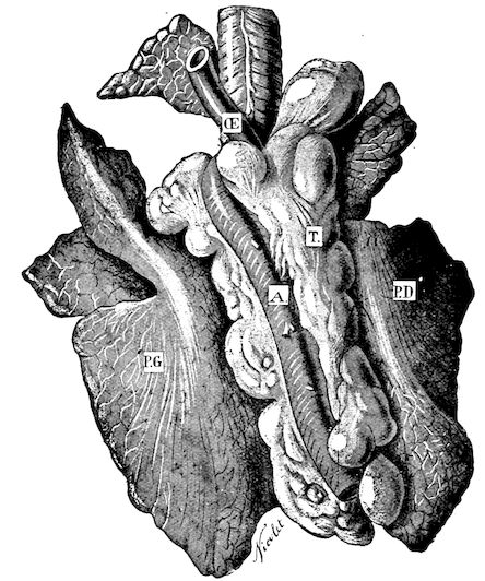
Fig. 278.—Well-developed tuberculosis of the mediastinal lymphatic glands. PG, Left lung; PD, right lung; Œ, œsophagus; A, posterior aorta; T, tuberculous lymphatic glands.
If, as often happens, the glands of the posterior mediastinum are affected and greatly enlarged, they may involve the œsophagus and the œsophageal nerves, interfere with deglutition and rumination, and thus produce marked disturbance. The animals only swallow with difficulty, and later rumination becomes impossible, the antiperistaltic movement not being powerful enough to overcome the resistance. Soon after eating, the patients exhibit tympanites, certainly only to a moderate extent, but the swelling is long in disappearing. This 699tympanites is due to the difficulty in eructation and to the impossibility of rumination. The contents of the rumen pass slowly towards the intestine until the onset of a fresh attack.
On account of digestive difficulties, the animals rapidly lose flesh, just as though they were suffering from extensive visceral lesions.
TUBERCULOSIS OF THE DIGESTIVE TRACT.
Tuberculosis of the digestive tract is rarer than tuberculosis of the lungs or lymphatic glands, and occurs in two well-differentiated clinical forms, that is to say, tuberculosis of the buccal and pharyngeal membrane and tuberculosis of the intestines. Tuberculosis of the liver, which is less easily recognised, is indicated by signs of dyspepsia.
Bucco-Pharyngeal Tuberculosis.—This may be primary or secondary, and occurs in the form of local or general glossitis or superficial ulcerative stomatitis.
In the former case the glossitis may be regarded as due to actinomycosis; in the latter it can only be mistaken for simple ulcerative stomatitis.
It is accompanied by difficulty in mastication, and still more, where the pharynx is invaded, in deglutition; abundant frothy salivation in feeding; sometimes by true spasm of the pharynx and rejection of masticated balls of food.
Locally the buccal mucous membrane (cheeks, tongue, pillars of the fauces, etc.) exhibit ulcerations, with festooned borders, in size something between a florin and a five-shilling piece, covered with a greyish-yellow, earthy-coloured exudation adhering firmly to the subjacent parts. The margin of the ulceration is but slightly indurated, and the tongue preserves its mobility, except in cases of general deep-seated glossitis.
The condition may continue for weeks and months without improvement.
Intestinal Tuberculosis.—Tuberculous enteritis is always accompanied by tuberculosis of the mesenteric glands and of the sublumbar lymphatic chain. It is indicated at first by chronic tympanites and peritonism, unaccompanied, however, by atony of the rumen; at a later period by intermittent diarrhœa, which attains a maximum and is followed by constipation. Finally, when there exist numerous intestinal ulcers, the diarrhœa is profuse and intractable, the animals rapidly grow exhausted, and the disease spreads to other organs with startling rapidity.
The food is ill-digested and the fæces have a repulsive odour, as has also the gas which escapes from the rumen when it is punctured.
700
TUBERCULOSIS OF THE GENITAL ORGANS.
In males, tuberculosis may attack the testicle and neighbouring organs and tissues; in females, the ovary, uterus, vagina, or udder.
Tuberculosis of the testicle is rare. Moussu appears only to have seen one case, namely, in the boar. The disease produces specific vaginitis and specific orchitis, the serous surfaces of the vaginal tunic becoming adherent, and tubercles forming in the depths of the testicle, whilst in time the testicle becomes the seat of fungoid growths.
Tuberculosis of neighbouring glands has only been observed in the case of the prostate. Clinically it is distinguished by symptoms which suggest difficulty in urination, and comprise frequent straining, efforts to micturate, dysuria, etc. Examination by the rectum reveals changes in the prostate, but affords no exact information as to their nature.
Tuberculosis of the genital tract in the female is usually a delayed complication of a preceding visceral tuberculosis, though it may occur as a primary disease, in which case the lesions are localised about the vulva or the vagina. Tuberculosis of the ovaries, Fallopian tubes, and uterus is much more common than that of the two organs just mentioned. It seems certain that the disease may be transmitted directly from the male to the female by copulation, in cases where the male animal has a lesion on the penis.
Tuberculosis of the vulva is rarer, and is indicated by swelling, sclerotic changes, and the presence of tuberculous nodules, varying in size between a lentil and a hazel-nut; after ulceration of these growths, a thick yellowish pus containing the specific bacilli is discharged.
Tuberculosis of the vagina is also indicated by hardening of the walls, sclerous infiltration, and the presence of deep-seated tuberculous nodules, which may or may not become ulcerated. It may follow uterine tuberculosis, the discharge from the uterus continually soiling and at length infecting the floor of the vagina. In such cases the lower wall of the vagina is thickened and infiltrated to a greater extent than the roof, and is sometimes intersected by transverse ulcerated suppurating folds. Bacteriological examination reveals the presence of bacilli.
Tuberculous invasion of the ovaries, Fallopian tubes, and uterus is externally indicated by signs of chronic metritis accompanied by a purulent discharge, which may or may not be fœtid, but always possesses special characteristics. The neck of the uterus is half open, and the discharge is continuous. The pus is of a greyish-yellow colour, ill-formed, grumous, or more frequently granular, and it sometimes accumulates in large quantities in the depressions of 701the vagina. Examination by means of the speculum is of great value in diagnosing such lesions. On examining the parts through the rectum, the walls of the uterus are found to be greatly thickened, sometimes indurated, bosselated or totally deformed.
The Fallopian tubes and ovaries may have attained enormous dimensions, and the normal anatomy of the parts is greatly altered both as regards dimensions and relations.
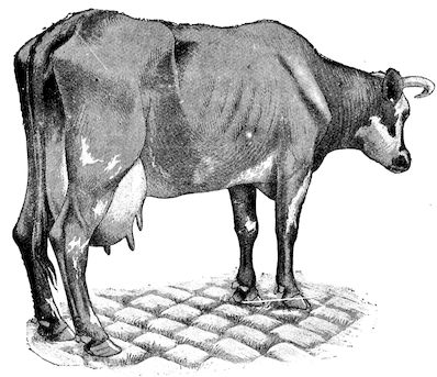
Fig. 279.—Hypertrophic tuberculous mammitis.
The almost inevitable consequences of tuberculosis of the genital organs are hypertrophy, induration or caseation of the subsacral and sublumbar lymphatic glands.
Udder.—Tuberculosis of the udder may be primary or secondary. When the infection is slight the results may escape notice for weeks or even months, the patients appearing to suffer only from subacute or chronic mammitis, while at the same time the milk preserves its ordinary appearance. In time, however, the mammitis becomes aggravated, the infected regions are enlarged, and the secretion becomes grumous, serous, curdled, and of a yellowish colour, afterwards ceasing altogether. In some cases one quarter only is attacked, though total mammitis is more common.
These forms of tuberculous mammitis tend towards hypertrophy, local hardening, and the formation of deeply-seated cavities containing pus, the gland itself sometimes acquiring enormous dimensions. The retro-mammary lymphatic glands are invaded even before the gland itself is seriously attacked. For a longer or shorter time the udder may externally appear healthy, although on manual examination these lymphatic glands are found to be indurated and bosselated.
TUBERCULOSIS OF BONES AND ARTICULATIONS.
Tuberculosis of the bones is seen only in young animals, and chiefly affects the vertebral column and the bones of the head. The 702limb bones are attacked as a rule only in the vicinity of diseased articulations.
The vertebral lesions corresponding to those in Pott’s disease in human beings are very difficult to discover before they produce complications, such as depression of the spine, compression of the spinal cord, paralysis, etc.
Lesions of the bones of the head or of the limbs are characterised by local deformity, destruction of osseous tissue, invasion of surrounding tissues, and by local symptoms peculiar to tumours originating in the periosteum.
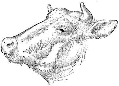
Fig. 280.—Perforating tuberculosis of the right frontal region.
Tuberculosis of joints produces special symptoms resembling those seen in the “white swellings” of man, that is, diffuse, œdematous, warm and moderately painful swelling of adjacent parts, accompanied by lameness of varying intensity. According to Guillebeau and Hess, many conditions described as strain or rheumatic arthritis are really tuberculous in character. They may remain stationary for a long time, or even recede under treatment. As a rule, however, these forms of tuberculous arthritis assume the fungoid type and prove incurable.
They are clinically distinguished from ordinary arthritis by the enormous swelling, which involves the extremities and a portion of the shafts of the bones. The adjacent muscles are chronically contracted, and the diseased joint is held semi-flexed. In course of time, if the patients are kept alive, abscess formation may occur, but this is seldom seen in practice, because the animals are slaughtered.
TUBERCULOSIS OF THE BRAIN.
Tuberculosis of the nervous centres, localised either in the meninges or the brain proper, may attack both young and old animals, not as a primary condition, but as a sequel to visceral disease, which, however, may have produced no outward indications, a fact that renders the diagnosis extremely difficult.
703When localised in the meninges, the disease produces the symptoms of ordinary meningitis, general weakness, vacillating, staggering or irregular gait, disturbed vision, variation in the size of the pupils, difficulty in swallowing, muscular twitching, cramp of the muscles along the upper margin of the neck, etc.
Tuberculosis of the brain proper seems more commonly to affect the anterior convolutions and the depths of the frontal and temporal lobes. It produces some of the symptoms of meningitis or symptoms suggestive of the existence of cœnurosis, as, for instance, walking in circles, lameness of central origin, without appreciable lesions of the limbs, prolonged kneeling, disturbed vision, generalised attacks of epilepsy or of Jackson’s epilepsy, spasm of the pharynx, general signs of compression of the brain or cerebral dropsy, dulness, coma, etc.
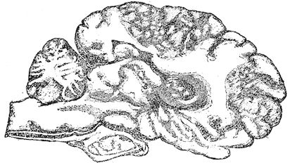
Fig. 281.—Tuberculosis of the brain. 1, 2 and 3, Tuberculous centres.
In the absence of pulmonary lesions it is extremely difficult to arrive at an exact diagnosis except after injection of tuberculin, for the above symptoms very closely resemble those of cœnurosis, brain tumours, and even tumours in the frontal sinuses.
TUBERCULOSIS OF THE SKIN.
Cutaneous tuberculosis is one of the rarest forms of the disease. It is distinguished by the formation beneath the skin of little hardened swellings varying in size between a hazel-nut and a walnut, and containing caseous or calcareous material. These swellings have no connection with the superficial lymphatic glands. They may be found grouped together within certain areas, or distributed irregularly over the whole body, particularly towards the base of the tail.
The condition may be mistaken for generalised sarcomatosis, from 704which, however, it is readily distinguished by microscopic examination of the contents of the swellings.
ACUTE TUBERCULOSIS—TUBERCULOUS SEPTICÆMIA.
However rapidly the above-described forms of tuberculosis may develop, the disease as a whole is always of long duration, and continues for months, or even for years. The development of these chronic forms may, however, be interrupted by various influences which cause it to assume an acute character, either for a time or continuously. Each intermittent attack aggravates the condition of the patient, but gradually subsides, with or without treatment. Continued attacks, however, rapidly lead to death; they may be seen in animals previously unsuspected of any grave disorder.
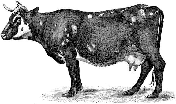
Fig. 282.—General appearance of a case of cutaneous tuberculosis.
The dominant symptom is continuous fever, accompanied by signs of disturbance of any or all of the chief bodily functions.
The temperature rises to 102° Fahr. (39° C.), or even to 104° or 105° Fahr. (40° or 41° C.), with morning and evening remissions of some hours. The respiration is accelerated. On auscultation it is often difficult to discover signs of chronic tuberculosis. The lung is the site of repeated congestive changes, resembling those of broncho-pneumonia or contagious pleuro-pneumonia. The pleura and walls of the chest become extremely sensitive, as in the last-named disease, and the abdomen may exhibit signs of peritonism, as at the beginning of acute peritonitis. The pulse rises to 80, 90, 100, or even 705120 beats per minute, and the urine contains albumen in notable quantities. This condition continues for weeks without apparent diminution, the patients refuse food, lose flesh with startling rapidity, and finally die of exhaustion.
It would be impossible from these peculiarities alone to identify the nature of the disease which causes such progressive organic wasting, as the continued presence of fever prevents the use of tuberculin, but fortunately the preliminary changes in the lungs, lymphatic glands, genital tract, etc., are sufficient in most cases for the purposes of diagnosis.
Sheep, Goats, and Pigs.—In the other domestic animals tuberculosis is only of secondary importance to the practitioner.
It has been seen in the sheep and goat, but almost exclusively as the result of experiment. It must be understood, however, that prolonged cohabitation with diseased oxen or lengthened sojourn in contaminated places may easily produce tuberculosis in the goat, though the sheep continues to resist for a somewhat longer period.
Clinically such tuberculosis presents little interest on account of its rarity.
The same remark applies to pigs; nevertheless, an entire herd may become infected, and it may be necessary, after making a preliminary post-mortem, to examine the other patients. All forms of the disease occur in pigs, the lung being most frequently affected, but tuberculosis also attacks the intestine, udder, lymphatic glands, joints, etc. The pig, in fact, is extremely susceptible to this disease, whilst the sheep is only subject to it in a comparatively trifling degree.
Diagnosis. The clinical manifestations of tuberculosis are so numerous and so various that it is often an extremely hard task to form a diagnosis. Without doubt detection is relatively easy in well-marked forms, such as tuberculosis of the lungs, lymphatic glands and genital apparatus, but even in such cases the symptoms must be reasonably well-marked.
At first, unless the lesions produce externally visible signs, diagnosis is impossible, and in the case of hidden forms, such as tuberculosis of the serous membranes, mediastinum, intestine, testicle, etc., all that can be done is to take into account the probabilities.
Clinical diagnosis is therefore possible, but only in exceptional cases can it be absolutely relied upon. Fortunately, methods of investigation increase and become more exact every day, so that the points which clinical examination is incapable of deciding are often cleared up in the laboratory. Bacteriological examination of morbid products, such as the nasal discharge, the products of suppuration, the milk or the diseased tissues, is a valuable means in many cases 706of determining the presence of the organism which causes the disturbance. In all cases, in fact, the tubercle bacillus should be sought for in order to confirm the diagnosis.
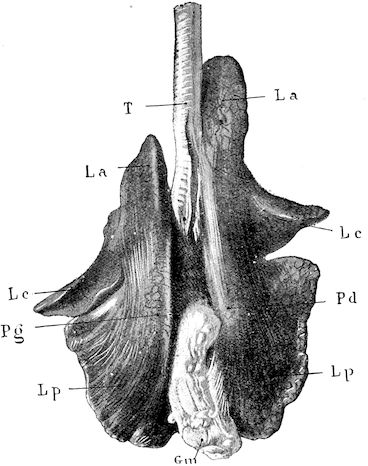
Fig. 283.—Tuberculosis of the posterior mediastinal lymphatic glands in a sheep which had been kept for two years in company with some tuberculous cows. PG, Left lung; PD, right lung; T, trachea; La, anterior lobes; L, middle cardiac lobes; Lp, posterior lobes; Gm, tuberculous and enlarged posterior mediastinal lymphatic glands.
If this method cannot be employed, as for example in tuberculosis of the liver, brain, etc., and the diagnosis is uncertain, the use of tuberculin constitutes the surest and easiest method of coming to a conclusion. Nocard’s researches have shown the precautions to be observed. A minimum febrile reaction of 2° Fahr. (1·5° C.) is, however, necessary before the existence of the disease can be affirmed.
Lastly, there remains a less rapid method, which aims at transmitting the disease to specially susceptible animals by inoculating with suspected materials, such as the nasal discharge, pus, milk or pulp of internal organs. This method is most valuable when bacteriological examination has failed and tuberculin has produced only doubtful results. The guinea-pig is the subject usually chosen, but some weeks, or even months, may elapse before definite results are obtained.
In those forms where ordinary methods of investigation prove sufficient it is well to bear in mind the symptoms which differentiate this disease from others closely resembling it.
Pulmonary tuberculosis, for example, should always be suspected whenever there exists frequent coughing, nasal discharge and poor bodily condition; if in addition to this respiration is found on auscultation to be rough, inspiration interrupted or rasping, expiration 707prolonged or blowing and the vesicular murmur non-existent, suspicion becomes almost a certainty. The diagnosis is even more assured if the respiration is of a blowing character at certain points, accompanied by snoring and sibilant râles and gurgling or cavernous souffles.
The distinction between pulmonary tuberculosis and chronic bronchitis or simple pulmonary emphysema is based on the increased resonance revealed by percussion in the latter case; the different character of expiration; the existence of a double breathing movement; the external appearance of the animal; the absence of bacilli from the nasal discharge, and the failure to react to tuberculin.
The condition cannot be mistaken for verminous broncho-pneumonia if the information furnished by auscultation and the results of microscopical examination of the discharge are taken into account, the eggs or embryos of strongyles being extremely easy to detect.
Tuberculosis of the pleura may be mistaken for peripneumonia if the observer trusts to percussion alone, but the auscultation sounds are then different, and an injection of tuberculin will remove any doubt.
Tuberculosis of the peritoneum is often suggested by the indications afforded by careful palpation (thickening of the walls of the abdomen, rigidity and sensitiveness), and is distinguished from ordinary acute peritonitis by the difference in the appearance of the animals and the absence of much fluid. Chronic exudative peritonitis and ascites also exhibit sufficiently well-marked characteristics to enable them to be differentiated from tuberculosis of the peritoneum, but this is certainly not true of adhesive peritonitis, and in cases of the latter kind tuberculin is the only means of confirming the diagnosis.
External tuberculosis of the retro-pharyngeal and cervical lymphatic glands resembles, at a first glance, simple inflammation of lymphatic glands, but in the last-mentioned disease the lesions are symmetrical and the glands still retain a certain amount of elasticity, whilst in tuberculosis they are bosselated, hard, and sometimes fluctuating.
Tuberculosis of the mediastinum is suggested by difficulty in swallowing, especially if tympanites follows soon after eating, eructation is absent, and rumination is arrested.
A careful study of the development of the ulcerations will also in most instances make clear the difference between tuberculous stomatitis and simple stomatitis or stomatitis due to actinomycosis. Intestinal tuberculosis and tuberculous enteritis are sufficiently characterised by persistent intractable diarrhœa accompanied by tympanites, 708by the fœtid character of the fæces, and by moderate but continued fever. Chronic diarrhœa, which alone presents some analogy with this condition, is never accompanied by permanent tympanites.
Tuberculosis of the genital organs in male animals is always liable to be mistaken for simple orchitis and the development of tumours in the testicle; an injection of tuberculin will, however, indicate the nature of the lesion.
When the symptoms of genital disease or chronic mammitis in female animals suggest that the disease is of a specific character, the diagnosis can frequently be confirmed by a microscopical examination of the pus or milk.
Finally, should the practitioner hesitate as to the nature of the lesions which are the cause of arthritis, deformity of bones, cerebral symptoms, etc., tuberculin again will in most cases settle the question.
The prognosis in cases of tuberculosis is extremely unfavourable, whatever the form of the disease or its manifestations. Clinically the disease should be regarded as incurable in the strict sense of the word, however limited may be the lesions. The affected animals are not all doomed to immediate death: some may be kept alive, and may even serve an economic purpose without necessarily endangering others; it is sometimes possible to fatten them, though the risks probably far outweigh the advantages, but one can never rely on recovery in any particular case.
The gravity of this disease is the greater inasmuch as it assumes so many forms, any one of which may result in the infection of other animals.
All those forms of the disease, such as tuberculosis of the respiratory, digestive and genital tracts, in which virulent material containing bacilli is discharged realise these conditions. The patient becomes a source of infection to others of its kind, a fact which more than anything else renders the disease so dangerous to the farmer and breeder.
Only in cases where the lesions are closed (as in tuberculosis of the lymphatic glands, serous membranes, joints, etc.) can the sufferers be regarded as innocuous, and—as these lesions are exceptional or at least, as animals suffering from them are very frequently afflicted with open lesions from which bacilli are continually being discharged—every tuberculous animal must be regarded from a clinical standpoint as a constant danger to its neighbours.
This, however, must not be understood to mean that there are not different degrees of danger. It is quite certain that a patient with pulmonary caverns which are constantly throwing off enormous 709quantities of material full of bacilli is much more dangerous than another suffering only from slight bronchial or tracheal lesions, though the danger in the latter case is none the less always present.
Treatment. There is no really curative treatment of tuberculosis.
It must not be thought, however, that we are completely helpless and that the present condition of affairs must be allowed to continue indefinitely. Nocard and Leclainche have minutely laid down the lines to be followed as regards prophylaxis, though unfortunately the measures recommended cannot always be carried out.
Tuberculin having been proved an exact means of detecting tuberculous lesions in animals even where none were suspected, it is desirable, firstly, to test all the animals in a given establishment with tuberculin; and, secondly, to separate into classes (1) all animals which have reacted, and (2) those which have resisted.
The stables, etc., should then be completely disinfected by sweeping, washing first with hot water, then with strong antiseptic solutions, brushing over the walls with quick-lime solution and fumigating with sulphurous acid or formic aldehyde. The healthy animals should then be placed in one shed and the diseased animals in another. In order to render this system of isolation really efficacious the isolated animals and the healthy animals should have nothing in common, and the persons tending the two classes of animals, the buckets and other utensils, the watering places, etc., should be kept rigorously apart.
The animals known to be tuberculous should as rapidly as possible be prepared for slaughter, and if pregnant cows are included in the number the calves should be removed to the healthy stable immediately after birth and brought up either on boiled milk or by a healthy mother, experience having shown that congenital tuberculosis is of rare occurrence.