Part 1 — SECTION XVII THE COMMUNICABLE DISEASES—PART I (continued 2)
Reading edition
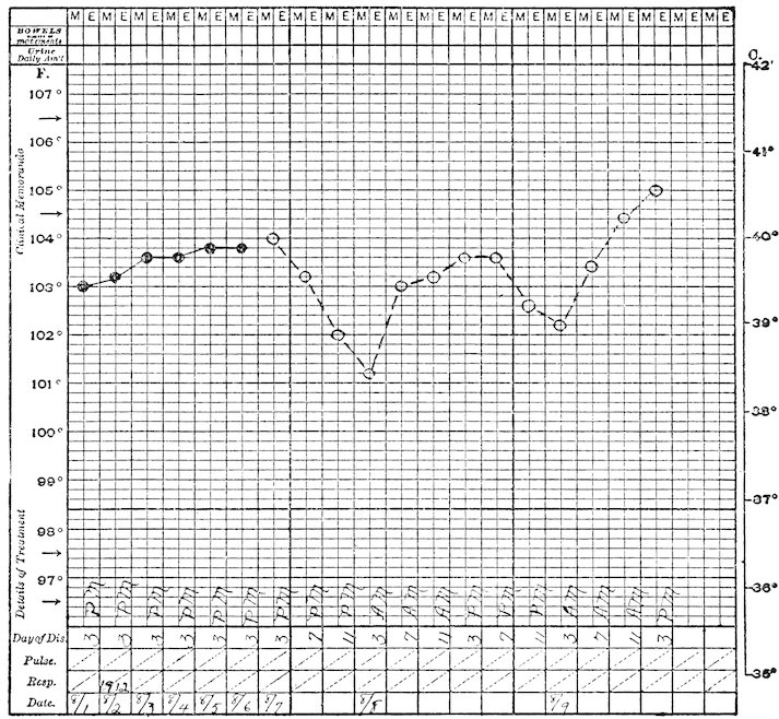
Chart Q. VARIEGATED CEBUS (Cebus variegatus). Tuberculous.
541
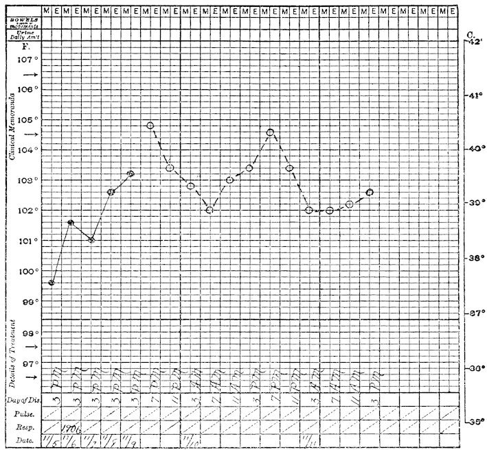
Chart R. SOOTY MANGABEY (Cercocebus fuliginosus). Tuberculous.
Another interesting experience concerns the exhibition of a group of Rhesus Macaques in an open “band stand” cage. The idea arose in an attempt to find a separate exhibition space for some good specimens that gave unsatisfactory charts, with the purpose of applying at the same time the “open air” treatment if tuberculosis existed. The experiment has been entirely successful since in the eleven years during which this enclosure has been used there has been but a single case of tuberculosis among twenty-six monkeys. Curiously enough this exception gave a good chart and we suspect it was a visitor infection; no secondary case arose from it. The animals housed in this cage keep in excellent condition, their coats responding to our severe winter by increasing in thickness and glossiness. Frozen toes, fingers and tails are sometimes seen but these monkeys seem just as happy as the others. Breeding is active and the young are lusty and husky. Practically the only deaths are due 542to accident, or to abuse of old and less vigorous members of the colony. We are unable to give comparative exhibition periods and death rates for monkeys in the large house and this open cage because some specimens have been changed from one to the other but it is certain that the appearance of the “band stand” monkeys is better than those in the house and there are four of eleven animals in the former which have been there eleven years and only four among the seventy in the exhibition house for that length of time.
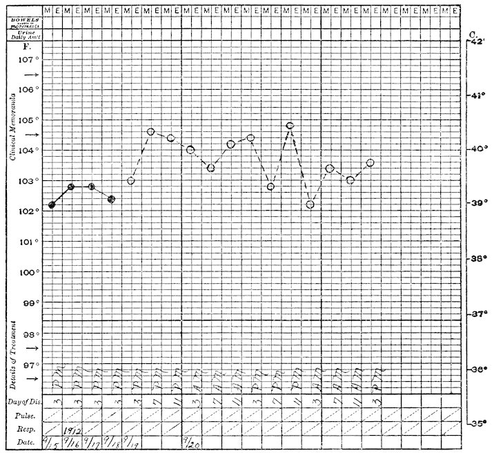
Chart S. VERVET MONKEY (Cercopithecus lalandii). Tuberculous.
543
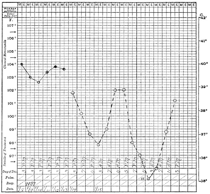
Chart T. GRIVET MONKEY (Cercopithecus sabæus). Tuberculous.
The results of the foregoing work seem to demonstrate that the tuberculin test permits the separation of tuberculous and non-tuberculous monkeys and that its employment serves the purpose of maintaining a healthy exhibition by excluding infected specimens. These experiences form further corroboration of the facts that tuberculosis begets tuberculosis, that a healthy individual is not a source of infection. It follows that an obviously tuberculous animal should not, need not, be a source of danger; the hidden or unrecognized case is the menace. There is little or no problem when an unequivocally good or bad temperature record is obtained; it is when there are slight variations from the standard for the group that decision as to the disposition of the specimen must be made. Nearly always such specimens are retested until the records are definite. If they be constantly irregular the animal is either sacrificed or exhibited in a separate cage far from other monkeys. It is by the sacrificing of 544infected specimens or the segregation of suspected ones that our collection is kept clear of disease.
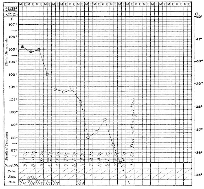
Chart U. CAMPBELL’S MONKEY (Cercopithecus campbelli). Non-tuberculous. (See page 533).
Hygiene of a general character must be maintained also. Our quarantine rooms are disinfected by formaldehyde and mechanical cleansing after every case detected as tuberculosis, and painted every two years. Monkeys associated with infected ones, are retested and then given a bath of carbolized water before being put on exhibition. The exhibition house is mechanically and chemically disinfected at the injection time each two years. All keepers are examined for tuberculosis upon beginning their employment and those handling monkeys, periodically thereafter. When a case of tuberculosis dies, all animals in 545the same and adjoining cages are removed for retest and the enclosure scrubbed and disinfected.
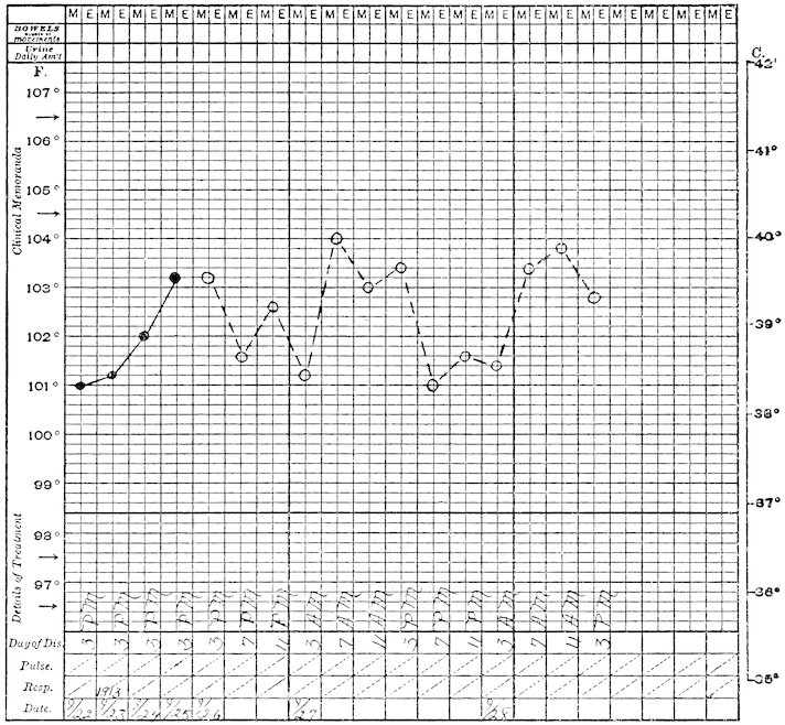
Chart V. WEEPER CEBUS (Cebus capucinus). Tuberculous.
There is a source of tuberculosis upon the importance of which we can only speculate—the visitor. There were two isolated cases in animals which had passed the test with unexceptionable charts, three and four months on exhibition; curiously enough no other cases occurred in their cages. These we have laid to visitor infection since no previous exposure can be traced for the specimens and no secondary cases occurred.
I can conclude this discussion of the tuberculin test and of the control of tuberculosis by its use, by mentioning the possibilities for the solution of the problem in man. While the eradication of the disease cannot be accomplished 546as easily as if a potential source could be eliminated by sacrifice, it will come in direct relation to the earliness of detection of infection and isolation of the sources of danger. Not so much the cough-racked consumptive but the unrecognized early lesion whose bearer hawks and spits in public places or at home, unaware of his malign power!
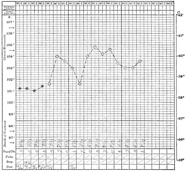
Chart W. CHACMA BABOON (Papio porcarius). Tuberculous.
The Skin and Eye Tests with Tuberculin.
The first of these can be dismissed briefly, for in a few cases it was absolutely of no value. A known tuberculous monkey was injected into the skin of the chest with 0.5 mg. of old tuberculin. The small bleb disappeared in a few hours and was followed by no reaction whatsoever. Other 547attempts likewise failed, some of them I believe due to the technical difficulty of injecting into the skin. This tissue is very thin, delicate and loose at the less hairy places where a reaction might be read—arm, chest, abdomen. The hairless parts of the rump might be used, but are so often scratched and soiled with dirt that readings might be misleading. The Von Pirquet test was done on the first mentioned specimen and was likewise negative. His tuberculin test was afterward positive.
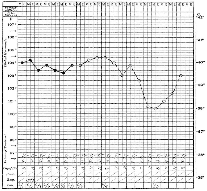
Chart X. BLACK APE (Cynopithecus niger). Tuberculous.
The ophthalmic reaction is highly spoken of in the New York Zoological Park and has been used elsewhere. It was tried by me at the time Doctor Blair first discussed it, but with variable results. One set of two monkeys was treated with Calmette’s purified tuberculin into the 548conjunctival sac and given a subcutaneous dose of old tuberculin. Another set received 1 per cent. old tuberculin into the conjunctivæ and the usual subcutaneous dose. Although all these monkeys gave a temperature reaction only one gave a conjunctival reaction. Fearing that the two tests simultaneously might be an unfair trial, another poor specimen was given an eye test which resulted negatively; a later subcutaneous test and autopsy revealed the disease. Because of these experiences and the fear that any reacting conjunctivæ might become secondarily infected from the uncertain personal hygiene of the beast, we decided to omit this method and rely upon the temperature test.
Pathological Effects of the Injection of Tuberculin.
It is generally believed that tuberculin injected into tuberculous animals, in doses large enough to produce a marked reaction at the site of disease, may stimulate the process to growth and spread and that certain parenchymatous organs in such bodies undergo degenerative changes. We can give little information concerning the first point because known infected animals have not been sacrificed during the test and we are not informed of the degree of morbid lesion in those dying, since we had no previous knowledge of its existence.
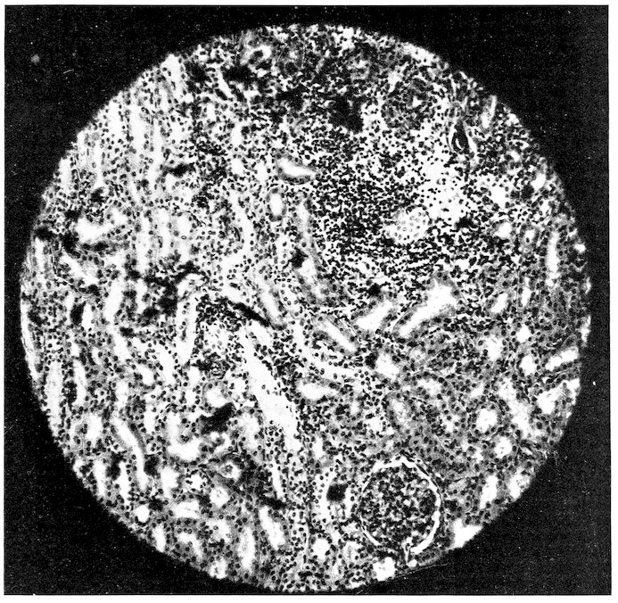
Fig. 58.—EFFECT OF TUBERCULIN ON THE KIDNEY. AN UNUSUALLY SEVERE REACTION IN THE RENAL TUBULES AND INTERSTITIAL TISSUE FOLLOWING A TEST DOSE OF TUBERCULIN. SUCH A MARKED CHANGE SUGGESTS THAT IN THIS CASE PRE-EXISTING RENAL DAMAGE WAS AGGRAVATED. THE ANIMAL WAS TUBERCULOUS BUT HAD NO LESIONS IN THE KIDNEY.
549An interesting and practically useful observation has, however, been made upon the kidneys of several monkeys dying shortly after tuberculin injection. It consists in a marked cloudy swelling of the renal epithelium and a congestion or even thrombosis of the glomerular capillaries, accompanied sometimes by increase of nuclei in the tuft and by amorphous material in the space of Bowman. Grossly such kidneys are but little changed, albeit the cortical zone may be dull and opaque and swell out slightly on section; very occasionally bloody streaks may separate the cortical and medullary striæ. In a few kidneys there have been suggestions of preëxisting nephritis but usually the findings are confined to those given above. At all events true glomerulonephritis is not often found. Monkeys which have this condition may or may not exhibit a behavior suggesting its existence. Sometimes it will be noted that the animal is dull and eats little, at other times the keeper will report that the cage is seldom wetted and we know of cases in which only an ounce or two of urine has been passed in a day. Two monkeys were distinctly ataxic and incoördinate and one of these had a convulsion. From one a specimen of urine showed albumen but no casts.
These signs of renal affection are not always alone nor are the kidneys necessarily the only part diseased since postmortem records show a variety of accompanying lesions, bronchitis and enteritis, for example. There are, however, several cases dying in a few days after tuberculin injection, both with and without tuberculosis, in which the renal changes were quite prominent; two examples, without tuberculosis, exhibited the damage to the kidneys very well and with no other evident visceral pathology. The relation of cause and effect may not be unequivocal, but these findings suggest that the condition of the kidneys deserves attention when tuberculin is to be injected. My associate, Dr. Corson-White, is firmly convinced that the substance whips up a preëxistent parenchymatous disease and wants to see a urinalysis from every monkey that is in any way abnormal.
The Tuberculin Test in Other Animals.
Two cases of tuberculosis occurred in White nosed Coatis (Nasua narica) so that it was decided to test their neighbors in the next cage. There is reproduced a composite (Y) of the temperature record of three of these animals after receiving 2 mg. of tuberculin under the skin. No tuberculosis was found in them at death, all dying 550within two years. The similarity to the primate type of temperature curve is striking.
Chart Z shows the course of temperature before and after tuberculin injection in a Bactrian Camel (Camelus bactrianus) ♀ which lived for some months and showed no infection at autopsy.
Charts AA and BB show the temperature ranges of respectively a healthy and a tuberculous Bison (Bison bison.)
Chart CC is that of a Malayan Sambur Deer (Cervus equinus) which died a week after injection, showing fibrocaseous tuberculosis. The failure to make an initial rise is noteworthy, but the fall in temperature may be explained by the severity of the lesions and the approaching death.
Chart DD represents daily and post-injection records of a Virginia Deer (Cervus virginianus) which at death was found free of the disease. Chart EE is that of a healthy American Elk (Cervus canadensis).
551
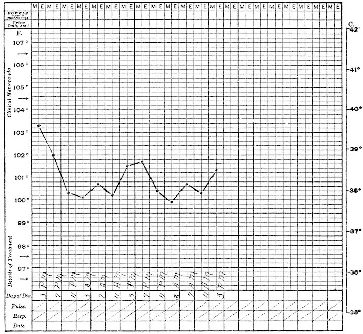
Chart Y. Composite chart of three non-tuberculous coatis.
552
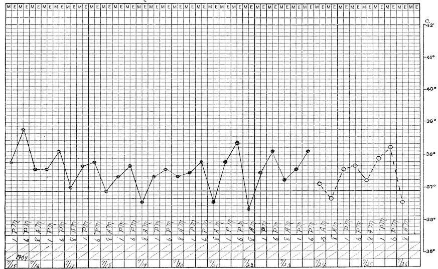
Chart Z. BACTRIAN CAMEL (Camelus bactrianus). Non-tuberculous.
553
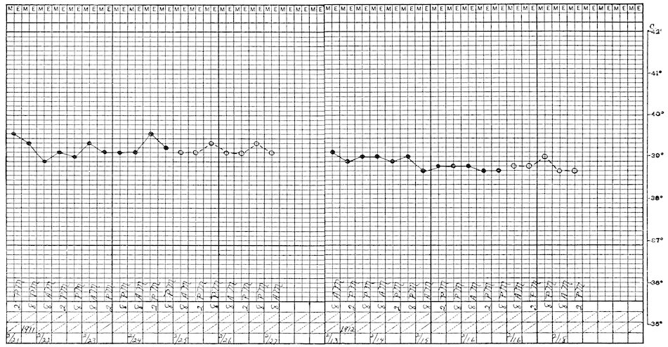
Chart AA. AMERICAN BISON (Bison bison). Non-tuberculous.
554
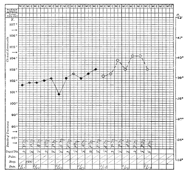
Chart BB. AMERICAN BISON (Bison bison). Tuberculous.
555
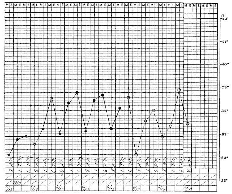
Chart CC. SAMBUR DEER (Cervus equinus). Tuberculous.
556
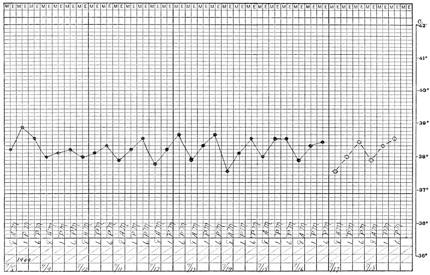
Chart DD. VIRGINIA DEER (Mazama virginiana). Non-tuberculous.
557
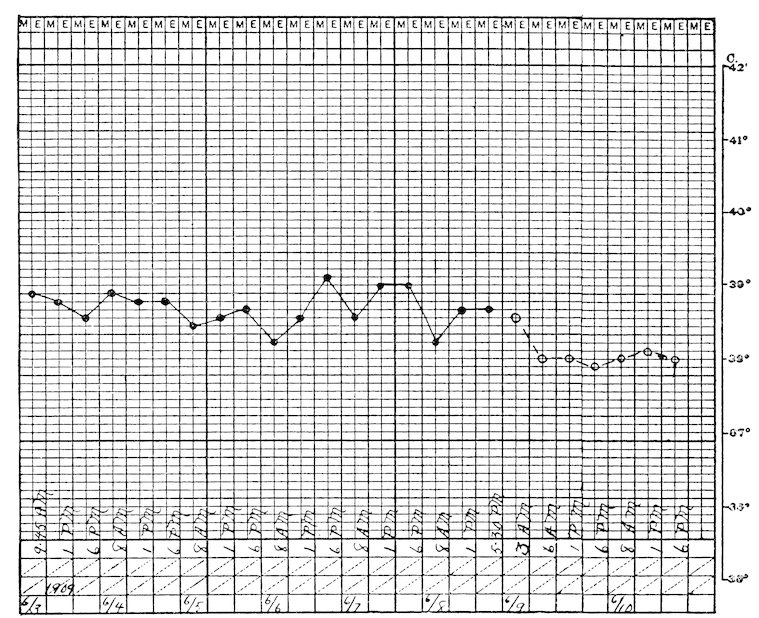
Chart EE. AMERICAN ELK (Cervus canadensis). Non-tuberculous.
558