DESCRIPTION OF THE MUSEUM AND ITS CONTENTS.
Reading edition
On entering the Museum, the visitor should bear in mind that the principal front faces the south, so that he will be looking due north, with the east on his right, and the west on his left hand.
It must also not be forgotten that a museum in a state of active growth is continually receiving additions as well as undergoing changes in the arrangement of its contents, and since these often occur faster than new editions of the Guide can be produced, there may be variations in the positions of some of the specimens from those here given.
The Central Hall.
Statues and Cases in the Central Hall.
On entering the hall the visitor will notice the bronze statue of the late Sir Richard Owen, K.C.B., Superintendent of the Natural History Departments of the British Museum (1856–1884). It is the work of Mr. T. Brock, R.A., and was placed in the Museum on March 17th, 1897. To the right of this is a marble statue of the late Professor T. H. Huxley, sculptured by Mr. E. Onslow Ford, R.A., which was unveiled on April 28th, 1900. In the first bay on the left is a bust, by Mr. Brock, of the late Sir W. H. Flower, Director of the Natural History Departments of the British Museum from 1884 to 1898. Most of the cases placed on the floor of the hall illustrate general laws or points of interest in natural history which do not come appropriately for illustration within the systematic collections of the departmental series.
Pigeons illustrating Variation under Domestication.
One group, in a case near the entrance to the hall, on the right, shows the great variation to which a species may become subject under the influence of domestication, as illustrated by examples of the best-marked breeds of Pigeons, all derived by selection from the wild Rock-Dove (Columba livia), specimens of which are shown at the top of the case.
Fowls and Canaries illustrating Variation under Domestication.
In the corresponding case on the left are further illustrations of the same subject. A pair of red Jungle Fowl (Gallus bankiva, or G. ferrugineus) of India represents the species from which the breeds of Domesticated Fowls are generally considered to have been derived. As examples of extreme modifications in opposite directions produced by selection, are exhibited the Japanese Long-tailed Fowls, in which some of the feathers (tail-coverts) attain a length of nine feet, and specimens of a breed in which the tail is absent. There is also shown a group of Fowls living wild in the woods of the Fiji Islands, which are descendants from domesticated birds introduced in the eighteenth century. A pair of Cochin Fowls is exhibited in the same case to display development in point of size and in the abundance of feathers on the limbs; while a pair of white “Silkies” illustrates a modification of the plumage, accompanied by a rudimentary condition of the tail-feathers. The pair of Coloured Dorkings exemplifies a breed cultivated in England.
A series of Canaries is shown in a case in the archway leading from the east side of the central hall to the north hall, as an example of a late addition to domesticated animals, these birds having been first imported into Europe from the Canary Islands in the early part of the sixteenth century. Specimens are exhibited of the wild birds, and of some of the modifications produced by selection.
Ruffs and Reeves, illustrating Changes of Plumage according to Sex and Season.
A case placed to the north of the one containing Fowls illustrates a remarkable instance of external differences in the two sexes and changes in plumage at different seasons, not under the influence of domestication. The birds in it belong to one species, the Ruff (Pavoncella, or Machetes, pugnax), of which the female is called Reeve; a member of the Plover family (Charadriidæ). In the upper division of the case are shown the eggs, newly-hatched young, and young males and females in the first autumn plumage; as well as adult males and females in winter, when both sexes are exactly alike in colour and distinguishable externally by size alone. The large group occupying the lower part of this case consists of adult birds in the plumage assumed in the breeding time (May and June). In the female the only alteration from the winter state is a darker and richer colouring, but in the males there is a special growth of elongated feathers about the head and neck, constituting the “ruff” from which the bird derives its name. In addition to this peculiarity, another, rare among wild animals, may be observed, namely, striking diversity of colour in different individuals. Of the twenty-three specimens shown no two are entirely alike.
Wild Ducks illustrating Seasonal Change of Plumage.
Next in order stands a case displaying the variations, according to season and age, in the plumage of the Wild Duck, or Mallard (Anas boscas). The most noticeable feature in the plumage-changes is the assumption in summer by males of an “eclipse-plumage,” resembling the one worn by females at all seasons. At other times the males are more brilliantly coloured than their partners. The eclipse-plumage corresponds to the winter, or non-breeding, dress of other birds which have a seasonal change.
Adaptation of Colour to surrounding Conditions.
On the same side of the hall follow two cases illustrating the adaptation of the colour of animals to their natural surroundings, by means of which they are rendered less conspicuous to their enemies or their prey. The first contains a specimen of a Mountain or Variable Hare (the common species of the north of Europe), a Stoat, and a Weasel, together with some Willow-Grouse and Ptarmigan, as well as an Arctic Fox, in their summer dresses. All were obtained in Norway, and show the general harmony of their colouring at this season with that of the rocks and plants among which they live. The second case displays examples of the same animals obtained from the same country in winter, when the ground was covered with snow. Such striking changes as these only occur in latitudes and localities where the differences between the general external conditions in the different seasons are extreme, where the snow disappears in summer and remains on the ground during most of the winter. Even some of the species here shown do not habitually turn white in the less severe winters of their southern range, as the Stoat in England and the Variable Hare in Ireland. A few permanent inhabitants of still more northern regions, where the snow remains throughout the year, such as the Polar Bear, Alaskan Bighorn Sheep, Greenland Falcon, and Snowy Owl, retain the white dress throughout the year. The whiteness of these animals must not be confounded with albinism, or whiteness occurring in individuals of species normally of a different colour, which is illustrated in a case on the other side of the hall.
Protective Resemblance of Desert Animals to their Surroundings.
The case on the east side of the hall nearest the great staircase contains examples of conformity of general style of colouring to surrounding conditions, as exemplified by some of the commoner Birds, Mammals, and Reptiles of the Egyptian desert, placed on the stones and sand amid which they habitually dwell. The advantage of this colouring in concealing the herbivorous species from their enemies, and in enabling the carnivorous to approach their prey unperceived, is obvious. Many excellent cases of concealment by adaptation to surroundings, especially in eggs and young Birds, may be seen among the groups in the Bird-gallery.
Mimicry.
More special modifications for the same purpose are shown in the adjacent bay on the east side of the hall by Insects which closely resemble the objects, such as leaves, twigs, etc., among which they dwell. The close imitation of a dead leaf, presented by the Leaf-Butterfly (Callima inachis), when its wings are closed, could not be surpassed. In a further stage of the same condition, called “Mimicry,” the object resembled, or mimicked, is another living animal, belonging to a different species, family, or even order. The resemblance in these instances is also believed to be for protection, or to be in some way advantageous to the animal in which it occurs. We know, however, so little of the habits and life-history of animals in a state of nature that many of the purposes supposed to be served by particular colours or appearances can only be regarded at present as conjectural. Whatever be the real explanation, the facts shown by the specimens in this bay are very curious, and worthy of careful consideration.
Group illustrating Albinism.
The next case on the east side of the middle of the hall contains a series of specimens illustrating albinism, a condition in which the pigment, or colouring matter, usually present in the skin, hair, or feathers, and giving the characteristic hue, is absent. Individuals in this condition occur among many animals of various kinds, and are called “albinos.” In some of the specimens shown in the case the albinism is complete, but in many it is partial, the absence of colouring matter being limited to certain portions of the surface. Other examples of complete or partial albinism are shown in the North Hall.
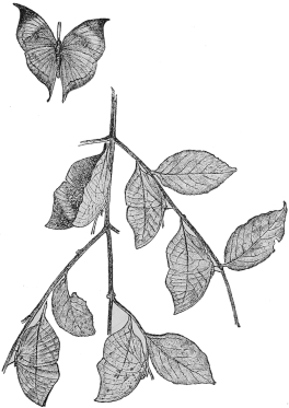
Fig. 1.—The Leaf-Butterfly (Callima inachis) in Flight and at Rest.
Group illustrating Melanism.
The adjacent case shows examples of the opposite condition called melanism, depending upon an excess of dark colouring matter or pigment in the skin and its appendages, such as hair, feathers, etc., beyond what is commonly met with in the species. This is by no means so frequent as albinism. A black Leopard in the middle of the case is a good illustration. This is not a distinct species, but merely an individual variety of the common Leopard, born from parents of the normal colour. A black Bullfinch is introduced as an example of acquired melanism, this bird having turned black in captivity.
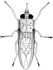
Fig. 2.—The Somali Tsetse-Fly (Glossina longipennis). Enlarged 4 diameters.
Shows the complete closure of the wings in the resting position, and the prominent proboscis, characteristic of the genus.
Group illustrating Intermediate Forms in Nature.
Another group shows that two forms of Crows which appear quite distinct, and, judged by their external characters, might be regarded as different species, may in a state of nature unite, and produce hybrid offspring. In the same case is exhibited a series of Goldfinches to show a complete gradation between birds of different colouring, which have been regarded as different species. Both these examples may by some naturalists be considered instances, not of the crossing of distinct species, but of “dimorphism,” or the occurrence of a single species in nature in two different phases. From whatever point of view they may be regarded, they illustrate the difficulty of defining and limiting the meaning of the term “species,” of such constant use in natural history.
Mosquitoes and Tsetse-Flies.
In the middle line of the hall are placed cases containing greatly enlarged models of certain Insects concerned in the spread of disease, such as Mosquitoes or Gnats (figs. 3 and 4), a House-Fly, Tsetse-Fly (fig. 2), and Plague-Flea; also still more enlarged gelatine models of mammalian blood-corpuscles, showing the parasites by which they are infested in the diseases respectively communicated by means of Mosquitoes and Tsetses. Models of the parasites themselves are also shown (figs. 5 and 6).
Malaria, or ague, is a disease caused by extremely minute parasites which live in the red corpuscles of the blood. Formerly malaria was believed to be contracted by merely breathing the air of marshy districts, but it is now proved that the parasites are transmitted from man to man by the “bite,” or rather “stab,” of a Mosquito or Gnat. The Common Mosquito or Stabbing Gnat (Culex pipiens), fig. 3, does not transmit the malaria-parasite; the Spot-winged Mosquitoes (fig. 4) of the genus Anopheles, abundant in England and nearly all parts of the world, being the carriers. The parasite multiplies not only in the human blood, but in the stomach and tissues of the Gnat—as shown in the models (fig. 5).
Tsetse-Flies are African blood-sucking Flies, with the mouth-parts adapted for piercing the skins of wild and domesticated mammals, human beings, and even crocodiles. The blood of some of the larger African animals is sometimes infected with a microscopic parasite (fig. 6), which, if sucked up by Tsetse-Flies when feeding, and subsequently introduced by them into the veins of domesticated animals such as horses and cattle, produces the fatal nagana, or Tsetse-Fly disease. Sleeping-sickness in man is caused in a similar manner, and is conveyed from infected to healthy individuals by two kinds of Tsetse-Fly, including that of which an enlarged model, 28 times (linear) natural size, is exhibited. The parasites and the red blood-corpuscles are enlarged 6,000 diameters.
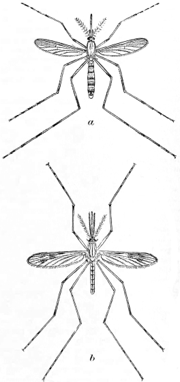
Fig. 3.—(a) The Common Gnat (Culex pipiens). Enlarged.
Fig. 4.—(b) The Spot-winged Mosquito (Anopheles maculipennis). Enlarged.
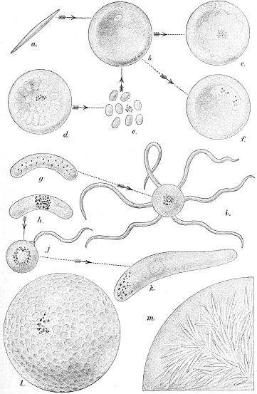
Fig. 5.—Life-History of the Malaria Parasite.
a, malarial germ, or sporozoite, as introduced into the blood by the mosquito; b, sporozoite after entry into blood-corpuscle; c, growth of sporozoite into an amœbula; d, division of amœbula to form merozoites; e, liberated merozoites; f, growth of merozoites into a crescent at expense of corpuscle; g, male, and h, female crescent; i, male cell with projections, which lengthen and are set free as spermatozoa; j, fertilisation of ovum by spermatozoon; k, fertilised egg as the active motile vermicule; sphere formed from the l, enlarged vermicule, after this has bored through the stomach-wall of the mosquito; m, segment of sphere at final stage of development, containing countless needle-shaped spores, which, when it bursts, escape as sporozoites into the organs of the mosquito’s body and pass through the salivary glands into the proboscis, and so infect a man bitten or pricked by the mosquito.
The House-Fly—see enlarged models of the perfect insect and its preliminary stages,—although not provided with a piercing proboscis, and consequently incapable of biting, sometimes plays an important part in spreading deadly diseases, such as cholera and enteric (typhoid) fever. In this case the disease-causing organisms are carried by the insect either in its intestine or adherent to the outer surface of its body, and thus House-Flies may spread disease by contaminating food.
Plague, which is a disease of rats and a few other rodents, especially the bobac Marmot, is communicated to man by the bite of the flea known as Xenopsylla cheopis, one of several species of fleas with which rats are infested. When a rat dies, the fleas that it has been harbouring seek another host, and may bite human beings, in which case, if the rat itself was suffering from the disease, an epidemic of plague may be the result. The model of the Plague-Flea exhibited is enlarged 200 times (linear).
The Arachnida, a group which includes the Spiders and can generally be distinguished from Insects by the number of their legs (four pairs instead of three pairs), include also the Ticks, which are responsible for the transmission of many deadly diseases which attack Man and Domestic animals. Enlarged models of a disease-carrying Tick are in course of preparation.
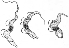
Fig. 6.
Trypanosoma gambiense, the parasite of Sleeping-sickness, very highly magnified. The occurrence of three kinds of individual, as shown in the figure, appears to be characteristic of this and certain other species of Trypanosoma.
(After Col. Sir D. Bruce.)
African Elephant.
In the middle line of the hall is placed a magnificent mounted skin of an African Elephant (Elephas africanus), fig. 7, from Rhodesia, standing about 11 feet 4 inches in height. The skull of the same individual is mounted on a stand below. Near by are exhibited three tusks, the largest of which measures 10 feet 2½ inches in length. On the north wall, on either side of the Darwin statue, are mounted two African Elephant heads.
Bays or Alcoves round the Hall.
Most of the bays or alcoves round the hall, five on each side, are (with the exception of the one at the north end of the right side) devoted to the Introductory or Elementary Collection, designed to illustrate the more important points in the structure of certain types of animal and plant life, and the terms used in describing them. This has been called the “Index Museum,” as it was thought at one time that it would form a sort of epitome or index of the general collections in the galleries. It is now mainly restricted to a display of the leading structural features in Vertebrated Animals and in Plants. The space being limited, the number of specimens is necessarily restricted. In examining this collection the visitor should follow each case in the usual order of reading a book, from left to right, and carefully study the printed explanatory labels, to which the specimens are intended to serve as illustrations.
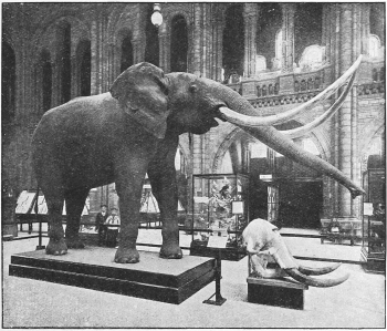
Fig. 7.—African Elephant (Elephas africanus).
The skull and tusks seen in the foreground are those of the stuffed specimen, the tusks in which have been modelled from another individual.
The bays on the west side (left-hand on entering the hall) are devoted to the Vertebrata, or Animals possessing a “backbone.” In Nos. I and II are shown the characters of the Mammals, which form the highest modification of this type; the wall-cases of No. I containing specimens of the bony framework (internal skeleton).
Bay I. Skeleton of Mammals.
In the first case (south side of the recess) may be seen a complete skeleton of a good example of the class—a Baboon Monkey, with the bones laid out on a tablet, and their names affixed. Below is a skeleton of the same animal articulated, or with the bones in their natural relation to each other, and also named. By examining these two specimens an idea may be obtained of the general framework of the bodies of animals of this class. In other parts of the case are exhibited various modifications of the skeleton to suit different conditions of life.
1. Man, showing a skeleton adapted for the upright posture.
2. A Bat, or flying Mammal, in which, by the great elongation of the fingers, the fore-limbs are converted into wings (fig. 8), supporting a web of skin stretched between them.
3. A Sloth, in which the tips of both limbs are reduced to mere hooks, by the aid of which the creature hangs back-downwards from the boughs of the trees among which it passes its entire existence.
4. The Baboon serves as an example of an animal walking on all four limbs in the “plantigrade” position, i.e., with the whole of the palms of the hands and the soles of the feet applied to the ground.
5. A small species of Antelope shows the characteristic form of a running animal, in which the limbs perform no office but that of supporting the body on the ground. This animal stands on the tips of the toes of its elongated, slender feet in the “digitigrade” fashion.
6. A Porpoise, adapted solely for swimming in the water. The fore-limbs are converted into flattened paddles, and the hind-limbs are entirely absent, their function being performed by the tail. The rudimentary pelvic bones are preserved.
The rest of the case is occupied by details of the skull in some of its principal modifications. At the top are diagrams of the structure of bone and cartilage as shown by the microscope.
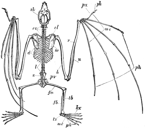
Fig. 8.—The Skeleton of a Flying-Fox, or Fox-Bat (Pteropus medius).
cl, clavicle; cv, cervical vertebræ; d, dorsal vertebræ; fb, fibula; fm, femur; h, humerus; hx, great toe, or hallux; l, lumbar vertebræ; mc, metacarpals; mt, metatarsals; ph, phalanges, or finger- and toe-bones; pv, pelvis; px, thumb, or pollex; r, radius; s, sacral vertebræ; sc, scapula; sk, skull; tb, tibia; ts, tarsus; u, ulna.
In the wall-case on the opposite (north) side of the bay the study of the skeleton of Mammals is continued by illustrations of the structure of the limbs. At the top of the case is a diagram showing the correspondence of the hand and the foot in their complete typical form, with the names applied to the different bones. The series of specimens below shows the principal deviations actually occurring from this typical condition, which, as may be seen, is very nearly preserved in the human hand. One series shows some of the stages of modification for special purpose (specialisation) by which a typical five-fingered hand becomes converted into the single-toed fore-foot of the Horse; while another series ends with the fore-foot of the Ruminants, sometimes, but erroneously, called a “cloven hoof,” in which only two toes remain. Similar changes are shown in the toes of the hind-foot, illustrating the same common plan running through infinite modifications in detail, enabling the organ to perform such a variety of purposes, and to exhibit such diversity of outward appearance. The existence of this common plan is now generally regarded as due to inheritance from a common ancestor.
Teeth of Mammals.
The central case of the bay contains a collection illustrating the principal characters of the teeth of Mammals. Its inspection should commence at the north-east corner, where the visitor will find himself after completing the survey of the specimens of skeletons in the wall-cases. In the first division are placed specimens showing the general characters of teeth, their form, the different tissues of which they are composed, the two great types of dentition in Mammals, homœodont and heterodont,5 the names and serial correspondence of the different teeth, and their development and succession. The principal modifications of teeth according to function are next shown by examples of forms adapted for fish-eating, flesh-eating, insect-eating, grass-eating, etc. The remainder of the case is taken up by examples of the dentition of the families of Mammals arranged in order, and prepared so as to display not only the shape of the crowns, but also the number and character of the roots by which they are implanted.
Bay II. Classification of Mammals.
In bay No. II the two wall-cases contain a collection arranged to show in a serial manner the orders and sub-orders of existing Mammals, by examples selected to illustrate the predominating characters by which these are distinguished. A brief popular account of the characteristics of the group, and a map showing its geographical distribution, are placed with each. This is intended to serve not only for an introduction to the study of the class by visitors to the museum, but also as a guide to a method of arrangement which may be adopted in smaller institutions.
Among the illustrations of the order Primates is placed the skeleton of a young Chimpanzee dissected by Dr. Tyson, which formed the subject of his work on the “Anatomy of a Pigmie,” published in 1699, the earliest scientific description of any Man-like Ape.
Skin of Mammals.
The central case of this bay contains illustrations of the outer covering or skin and its modifications in the class of Mammals, divided into the following sections:
1. Expansion of skin to aid in locomotion, as the webs between the fingers of swimming and flying animals, the parachutes of flying animals.
2. The development of bony plates in the skin, found among Mammals only in the Armadillos and their allies. The cast of a section of the tail of a gigantic extinct species (Glyptodon) shows a bony external as well as an internal skeleton.
3. The outer covering modified into true scales, much resembling in structure the nails of the human hand. This occurs in only one family of Mammals, the Pangolins, or Manidæ.
4. Hair in various forms, including bristles and spines. The two kinds of hair composing the external clothing of most Mammals, the long, stiffer outer hair, and the short, soft under-fur, are shown by various examples.
5. The special epidermal appendages found in nearly all Mammals on the ends of the fingers and toes, called according to the various forms they assume, nails, claws, or hoofs.
6. The one or two unpaired horns of the Rhinoceroses, shown by sections to consist of a solid mass of hair-like epidermic fibres.
7. The horns of Oxen, Goats, and Antelopes, each consisting of a hollow conical sheath of horn, covering a permanent projection of the frontal bone (the horn-core).
8. The antlers of Deer, forming solid, bony, and generally branched projections, covered during growth with soft hairy skin, and in most cases shed and renewed annually.
On the wall is arranged a series of antlers of an individual Stag or Red Deer (Cervus elaphus), grown and shed (except the last) in thirteen successive years, showing the changes which took place in their size and form, and the development of the branches, or tines, in each year. In old age the number of these tines tends to diminish.
On the north side of the table-case are shown dissections of the principal internal organs of Mammals.
Bay III. General structure of Birds.
Bay No. III is devoted to the class of Birds. An Albatross (Diomedea exulans) mounted with the wings expanded shows the most important characters by which a Bird is externally distinguished from other animals. The body is clothed with feathers, which (in the majority of Birds), by their great size and special arrangement upon the fore-limbs, enable these to act as organs of flight. The mouth is in the form of a horny beak. A nestling Albatross shows that at this stage of its existence the bird is not clothed with ordinary feathers, but with soft down, which serves to keep the body warm, although it confers no power of flight. An Emu and an Apteryx in the lower compartment of the case display the exceptional condition (found only in a comparatively few members of the class) of Birds with wings so small as to be concealed beneath the general feathery covering of the body, and quite useless. In the Penguins, of which two species are shown in the case, the wings are reduced to the condition of fins, and are serviceable only for progress through water.
In the first wall-case the principal features of the skeleton of the class are shown. Sections of bones exhibit the large air-cavities within; a complete skeleton of an Eagle, with the bones separated and named, and mounted skeletons of the Ostrich, Penguin, Pelican, Vulture, Night-Parrot, Fowl, etc., show the chief modifications of the skeleton. The Apteryx possesses the smallest, and the Frigate-bird the longest bones of the wing, the correspondence of which can be readily traced by means of the labels attached to them. The under surfaces of the skulls of various birds are shown with the different bones coloured to indicate their limits and relations; these are followed by a series of the different types of sternum or breast-bone.
The second wall-case contains further illustrations of the anatomy of Birds. In the left-hand part a series of wings of Birds displays the form characteristic of different groups; while above them are a few of the different types of tails, supplementing the series of tails in the table-case. Very instructive is a series of skins of white chickens of the same brood at different ages, displaying the gradual replacement of the down by the adult plumage.
The table-case in the middle of the bay contains illustrations of the external characters, the beak, the feathers, and the tail, as well as of the fore and hind limbs, or wings and feet. By the aid of the explanatory labels, the essential characters and the principal modifications of all these parts may easily be followed.
Two cases on the wall in the vestibule leading to the Fish Gallery illustrate the chief modifications of the eggs of Birds, and their differences in structure, number, form, size, texture of surface, and colour. On the side of the main staircase opposite are specimens illustrating the parasitic nesting habits of certain Cuckoos and various other Birds; while near by is a remarkably fine series of the eggs of Cuckoos with those of the Birds among which they were respectively deposited. On the opposite (east) side of the staircase the visitor will find a case showing the remarkable variation in colouring and markings displayed by the eggs of the Guillemot.
Bay IV. General structure of Reptiles and Amphibians.
The fourth bay on the west side of the hall exhibits the leading peculiarities in the structure of Reptiles and Amphibians. Owing to the large number of groups in the former class now extinct, many fossil specimens, or plaster reproductions of the same, are shown. The wall-case on the south side of this bay illustrates the different ordinal groups of Reptiles—living and extinct. Very instructive are the skeletons of Tortoises and Turtles, showing the relations of the vertebræ and limb-bones to the bony part of the shell. Lizards and Snakes are mostly represented by coloured casts. The extinct Dinosaurs are represented by a small-sized model of Iguanodon, together with a photograph of the skeleton and a plaster-cast of the bones of the hind-foot showing the three toes.
The adjacent side of the table-case shows the modifications of the backbone, or vertebral column, of the ribs, and of the limbs, in the different groups of the class. Specially noticeable are examples of five types of Skink-like Lizards, exhibiting the gradual diminution in the size of the limbs and their final disappearance.
The opposite, or north, side of the table-case displays the different modifications of the skull and teeth of living and extinct Reptiles. In some, like Crocodiles and Ichthyosaurs, the jaws are armed with a full series of sharply pointed teeth, while in others, like the Tortoises and Turtles, they are devoid of teeth and encased in horn. Very remarkable is the approximation to a carnivorous mammalian type presented by the dentition of some of the extinct mammal-like Reptiles, or Theromorphs, and equally noticeable are the palatal crushing teeth of certain other extinct Reptiles known as Placodus and Cyamodus. The peculiar dentition of the New Zealand Tuatera, and likewise that of its extinct European and Indian ally Hyperodapedon (fig. 9), are also shown.
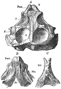
Fig. 9.—Skull of the Giant Tuatera (Hyperodapedon gordoni), from the Triassic Sandstone of Lossiemouth, Elgin, (¼ nat. size). A, upper surface of skull; B, palatal aspect of skull; C, under side of front of lower jaw; Pmx, premaxillary bone; Mx, maxillary; Pl, palatal teeth; Md, lower jaw; O, orbit, or eye-socket; N, nostrils; S, temporal pit; S’, lateral temporal fossa.
The brain and other internal organs of Reptiles are displayed in the left half of the wall-case on the north side of this bay, in which are also shown the eggs of many species, in some cases with the embryo.
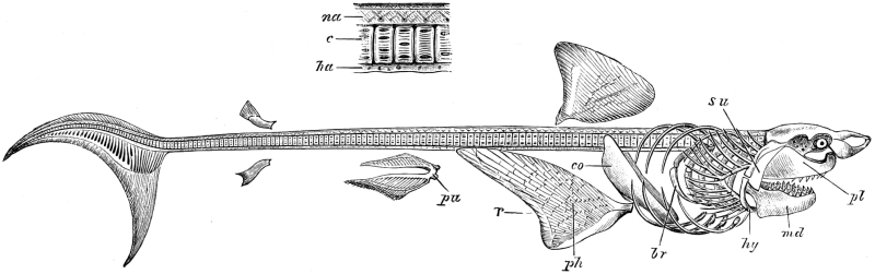
Fig. 10.—Skeleton of the Great Blue Shark (Carcharodon rondeletii), with portion of backbone on a large scale. pl, functional upper jaw, and su, its reflected portion; md, lower jaw; hy, ceratohyal; br, branchial arches; co, pectoral girdle; ph, cartilaginous portion of pectoral, or front paired fin; r, dermal portion of pectoral fin; pu, pelvic, or hind paired, fin; c, centra, or bodies, of the vertebræ; na, neural, or upper, and ha, hæmal, or lower, arch. The median fins are not lettered.
In the right half of the same case are exhibited a number of preparations showing the external form and internal structure of Frogs and Salamanders, or Amphibians, living and extinct. The Giant Salamander of Japan (Megalobatrachus or Cryptobranchus) is represented by a stuffed specimen; but the Newts, Salamanders, and Frogs are shown in spirit. Very curious is the almost colourless and blind Olm (Proteus) from the caves of Carniola; as also are the so-called Cœcilians, or Apoda, which have the habits and, in some degree, the appearance of large worms. Special specimens exhibit the structure of the extinct Labyrinthodonts, in which the hinder half of the skull is completely roofed over by bone; while the teeth in many instances exhibit a curious in-folded arrangement from which the group derives its name.
Bay V. Structure of Fishes.
The last bay (No. V) on the west side of the Central Hall is devoted to the display of the form and structure of Fishes.
The wall-case on the left side of this bay exhibits the external form of several characteristic types of Fishes, such as the Pike, Cod, Turbot, Dog-fish, and Skate, with the names of the various fins affixed. A striking specimen is the skeleton—mainly cartilaginous—of the Great Blue Shark (Carcharodon rondeletii), fig. 10, which occupies the greater portion of this case. It should be noted that, as in all Sharks and Rays, the upper jaw does not correspond with that of the higher Vertebrates; and particular attention should be devoted to the structure and arrangement of the arches supporting the gills.
In the south side of the table-case in this bay are shown a number of dissections, mounted in spirit, displaying the different types of skeletal structure presented by the fins in various groups of Fishes. One of the most remarkable of these types occurs in Ceratodus forsteri, the Queensland Lung-fish, in which the skeleton of the fin consists of a central jointed rod, from each side of which diverge narrower jointed rods. Alongside are specimens showing special modifications of certain fins, as in the Flying Fish (fig. 11) and Flying Gurnard (fig. 12), for the purpose of sustaining the body in the air, or, as in Pentanemus, to serve as organs of touch. Specimens of the West Indian Goby and the Lump-Sucker show modifications of the pelvic fins in connection with a sucker on the lower surface of the body; while other preparations display the pectoral (Doras) and pelvic fins (Monocentris) reduced to the condition of saw-like spines.
The structure of the skull of Fishes is illustrated in another part of the same side of this case. From this the visitor may learn how the primitive cartilaginous skull of the Sharks (fig. 10), Rays, Chimæras, and Lung-fishes has been gradually modified, by the addition of superficial sheathing-bones, into the bony skull of modern Fishes, such as the Cod and Perch.
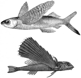
Fig. 11.—The Flying Fish (Exocœtus).
Fig. 12.—The Flying Gurnard (Dactylopterus).
The north side of the table-case in bay V is mainly devoted to the display of the different types of scales, spines, and teeth found among Fishes. In one corner are the enamelled “ganoid” scales of the modern American Bony Pike (Lepidosteus) and the African Bichir (Polypterus) alongside those of certain extinct forms. A scale of the Tarpon, or King-of-the-Herrings, illustrates the largest development in point of size of the modern “cycloid” type. Spines of the Porcupine-fish show an extreme development of this kind of structure. Diagrams and spirit-preparations illustrate the mode of attachment and succession of fish-teeth. A large series of the teeth of Sharks and Rays displays the gradual passage from those of the ordinary pointed form to others arranged in a pavement-like manner and adapted solely for crushing. Both types occur in the Port Jackson Shark (fig. 13), but those of some Rays are solely of the pavement modification. Very remarkable is the dental structure in the Parrot-fish. The west end of this side of the case shows the various modifications assumed by the teeth of the modern Bony Fishes; among which, as exemplified by the Wrasse, teeth are developed on the bones of the throat, as well as on those of the jaws. Throughout this case specimens, or models, of the teeth of extinct Fishes are placed side by side with those of their nearest living relatives.
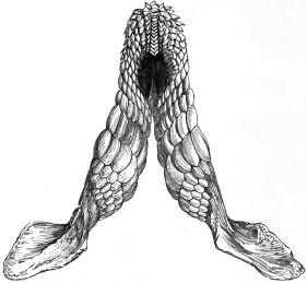
Fig. 13.—A Jaw of the Port Jackson Shark (Cestracion philippi), showing sharp teeth in front and crushing ones behind.
The wall-case on the north side of this bay shows the history of the development of various Fishes, together with the form and structure of the gills, brain, heart, digestive system, and other organs.
Lancelet.
A small case affixed to the pillar at the entrance of the fifth bay illustrates the structure of the Lancelet (Branchiostoma, or Amphioxus), by the aid of spirit-specimens, enlarged models, and coloured diagrams. One of the most remarkable features in the structure of this strange and primitive little creature is the outer cavity enclosing the part of the body which contains the large and complex pharynx. The Lancelet was formerly included among the Fishes, but is now accorded the rank of a class (Cephalochorda) to itself.
Leaving bay VI, next the principal staircase on the east side of the central hall, which is devoted to illustrations of heredity, especially in relation to the Mendelian theory, and to modes of Flight in Vertebrates and Insects, we pass on to a table-case assigned to the illustration of “Mimicry” and kindred phenomena. Most of the examples shown occur among Insects; but one example among Mammals and a second in Birds are illustrated. Very striking is a coloured sketch showing a group of red and black caterpillars from Singapore grouped side by side on the stem of a plant so as to present a remarkable similarity to a succulent fruit.
Bay VIII.
Bay IX.
In bay VIII, on the eastern side of the central hall, is displayed an exhibition illustrating trees, native to or grown in Britain. The winter and summer states are indicated by photographs, and the foliage, flowers, fruits, seedlings, and texture of wood and bark by specimens, models, and drawings. Bays IX and X are intended to illustrate the general characters of the great groups of the Vegetable Kingdom. Bay IX, in course of arrangement, is devoted to the Cryptogams (Ferns, Mosses, Fungi, Seaweeds, and Lichens).
At the back of the bay is a fine polished section of a buttress from the base of the Tapang (Abauria excelsa), the largest tree in Borneo, which attains a height of 250 feet.
Bay X. Seed-bearing Plants.
The last bay (No. X) is devoted to the Seed-bearing Plants, which are characterised by the formation of a seed—the result of the fertilisation of an ovule by the male cell which is developed in the pollen. The series begins on the left hand side with the Pteridosperms, an extinct group combining the characters of Ferns and Seed-plants and forming a link between them. Then follow the Gymnosperms (Cycads, Pines, Firs, etc.), in which the seed is borne naked on an open scale which generally forms, with others like it, the characteristic cone. Certain points in the development of pollen and ovule recall similar stages in the Fern group, and indicate that the Gymnosperms stand nearer to the Cryptogams than do the Angiosperms, the other and larger group of Seed-plants. The Gymnosperms are also the older group, and contain many extinct forms. In the Angiosperms the seed is enclosed in the fruit, and in the development of pollen and ovule almost all traces of a cryptogamic ancestry have been lost; the great development of the flower is a characteristic feature of the Angiosperms. The arrangement of the vegetative parts of the plant is based on its separation into root, stem, and leaf. In the right-hand wall-case the upper series of specimens illustrates the leaf, its form, veining, direction, the characters of its stalk and stipules, its modification for special purposes, and its arrangement on the stem and in the bud. Below, the stem and root are similarly treated, and above are some anatomical drawings. The display of the root is continued in the lower part of the opposite wall-case. In the central case the chief types of the flower with its parts, the fruit, and the seed are exhibited.
At the back of the bay is a large transverse section of the Karri tree (Eucalyptus diversicolor) of Western Australia, a species which grows to a height of 400 feet. The tree from which the section was cut was about 200 years old when felled.
The Introductory Collection of Minerals will be found in the gallery devoted to the Mineral Department (see p. 90).
The North Hall.
Domesticated Animals,6 Hybrids, and Economic Zoology.
The North Hall, or that portion of the building situated to the northward of the principal staircase, is used for the exhibition of the more important breeds of Domesticated Animals, as well as of examples of Hybrids and other Abnormalities. A series of specimens illustrative of Economic Zoology is likewise temporarily placed here.
The examples of Domesticated Mammals include Horses, Cattle, Sheep, Goats, Llamas, Dogs, Cats, and Rabbits. One of the main objects of this series is to show the leading characteristics of the well-established breeds, both British and foreign. In addition to Domesticated Animals properly so called, there are also exhibited examples of what may be termed Semi-domesticated Animals, such as white or parti-coloured Rats and Mice.
The skulls and skeletons of celebrated Horses7 of all breeds, including those of the Thoroughbreds “Persimmon” (presented by His Majesty King Edward VII.), “Stockwell,” “Bend Or,” and “Ormonde,” and of the Shire “Blaisdon Conqueror,” form a notable feature of the series. In another case is exhibited the dentition of the Horse at different periods of existence; while on the opposite side of the same is illustrated the evolution of the Horse from three-toed and four-toed ancestors, and also certain peculiarities distinguishing the skulls of Thoroughbreds and Arabs from those of most other breeds.
Among the more notable exhibits are a mounted specimen of a Spanish Fighting Bull, which belongs to an altogether peculiar breed, and heads of Spanish Draught Cattle, presented by H.M. King Edward VII. Among the Sheep, attention may be directed to the four-horned, fat-tailed, and fat-rumped breeds, and also to the small breed from the island of Soa, as well as the curious spiral horned Wallachian Sheep. The so-called wild cattle of Chillingham Park are included in this series, since they are not truly wild animals, but are descended from a domesticated breed. The celebrated Greyhound “Fullerton” is shown among the series of Dogs, which also comprises examples of the Afghan Greyhound, and of the Slughi or Arab Greyhound. Small-sized models of Cattle, Horses, Sheep, and Pigs also form a feature of the series.
A hybrid between the Zebra and the Ass is shown in one of the cases; while photographs illustrate the results of experiments undertaken by Professor Ewart in cross-breeding between Burchell’s Zebra and the Horse. An example of the Lion-Tiger hybrids born many years ago in Atkins’ menagerie is likewise shown.
A fine series of hybrid Ducks and hybrid Pheasants is exhibited in the north hall.
Skeletons of Man and Horse.
Facing the visitor as he approaches the middle of the north hall are the skeletons of a Man and of a Horse, arranged for comparison with each other, and also to show the position of the bones of both in relation to the external surface. In the case of the Horse, the skin of the same animal from which the skeleton was prepared was carefully mounted, and, when dry, divided in the middle line; one half, lined with velvet, has been placed behind the skeleton. In the case of the Man, the external surface is shown by a papier-maché model, similarly lined and placed in a corresponding position. As all the principal bones of both skeletons have their names attached, a study of this group will not only afford a lesson in comparative anatomy, but be of practical utility to the artist.
Section of “Big Tree.”
Against the wall dividing the north hall from the central hall is placed a section of a very large Wellingtonia or “Big Tree” (Sequoia gigantea), which was cut down in 1892 near Fresno, in California. It is about fifteen feet in diameter, and perfectly sound to the centre, showing distinctly 1,335 rings of annual growth, which afford exact evidence of the age of the tree. An instantaneous photograph, taken while the tree was being felled, is placed near by, and shows its general appearance when living. The height of the tree was 276 feet.
The exhibits of Economic Zoology at present occupy the northern division of this hall. In the western wall-case are specimens showing the injuries caused to trees by various insects. The table-cases contain examples of the damage done in Britain to fruit, roots, corn, and garden and vegetable produce, with specimens of the insects, and hints as to methods of destruction. There are also examples of injury done by insects abroad to cotton, tea, coffee, etc. In the cases under the windows are various parasites affecting man and domesticated animals.
Staircase and Corridors.
Statue of Darwin.
On the first landing of the great staircase, facing the centre of the hall, is placed the seated marble statue of Charles Darwin (b. 1809, d. 1882), to whose labours the study of natural history owes so vast an impulse. The statue was executed by Sir J. E. Boehm, R.A., as part of the “Darwin Memorial” raised by public subscription. It was unveiled and placed under the care of the Trustees of the Museum on the 9th of June, 1885, when an address was delivered on behalf of the Memorial Committee by the late Professor Huxley, P.R.S., to which His late Majesty King Edward VII. (then Prince of Wales), as representing the Trustees, replied.
Statue of Banks.
Above the first landing the staircase divides into two flights, each leading to one of the corridors which flank the west and east sides of the hall and give access to the galleries of the first floor of the building. Near the southern ends of these corridors two staircases join to form a central flight leading to the second floor. On the landing at the top is a marble statue by Chantrey of Sir Joseph Banks (b. 1743, d. 1820), who for 41 years presided over the Royal Society and was Trustee of the Museum. His botanical collections are preserved in the adjoining gallery, but his library of works on natural history, also bequeathed to the Museum, remains at Bloomsbury, where the statue, erected by public subscription in 1826, stood until it was removed to its present situation in 1886. On the wall above is displayed a series of unusually fine heads of Indian Big Game Animals, bequeathed by Mr. A. O. Hume, C.B., in 1912.
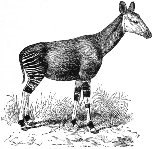
Fig. 14.—A Female Okapi (Okapia johnstoni).
African Antelopes.
The west, south, and east corridors contain a portion of the collection of mounted Mammals for which there is not room in the gallery immediately adjoining. The specimens placed here include a large number of species of the finest African Antelopes, animals remarkable for their beauty, for their former countless numbers, and for their threatened extermination in consequence of the inroads of civilized man into their domain.
Giraffes and Okapi.
In a case at the head of the staircase leading to the east corridor are several mounted specimens of Giraffes, and near by a skeleton of the same. Alongside the former is placed a case containing the heads and necks, together with skulls, of the various local races of Giraffes; while in a third are displayed three specimens of their near ally the Okapi (fig. 14) of the Congo Forest, as well as a skeleton of the same.
Collections of Humming-Birds.
The collection of Humming-Birds (Trochilidæ) arranged and mounted by the late Mr. John Gould, and purchased for the Museum in 1881, is principally shown in the vestibule leading from the hall to the Fish-gallery, but a few cases are placed on the pillars of the staircase. Another large collection of these birds, presented in 1913 by Mr. E. J. Balston, of Maidstone, is exhibited in the corridor leading to the Whale-room.
WEST WING.
The whole of the west wing of the building is devoted to the collections of recent Zoology.
(A) Ground Floor.
Bird Gallery.8
The ground floor is entered from the west side (left hand) of the central hall, near the main entrance of the building. The long gallery, extending the entire length of the front of the wing as far as the west pavilion, is assigned to the exhibited collection of Birds, the study-series of the same group being kept in cabinets in a room behind.
Systematic Series in Wall-cases.
The wall-cases contain mounted specimens of all the principal genera, placed in systematic order, beginning with the Crows and Birds of Paradise on the left hand on entering, and ending with the Ostriches, Emus, etc., on the right.
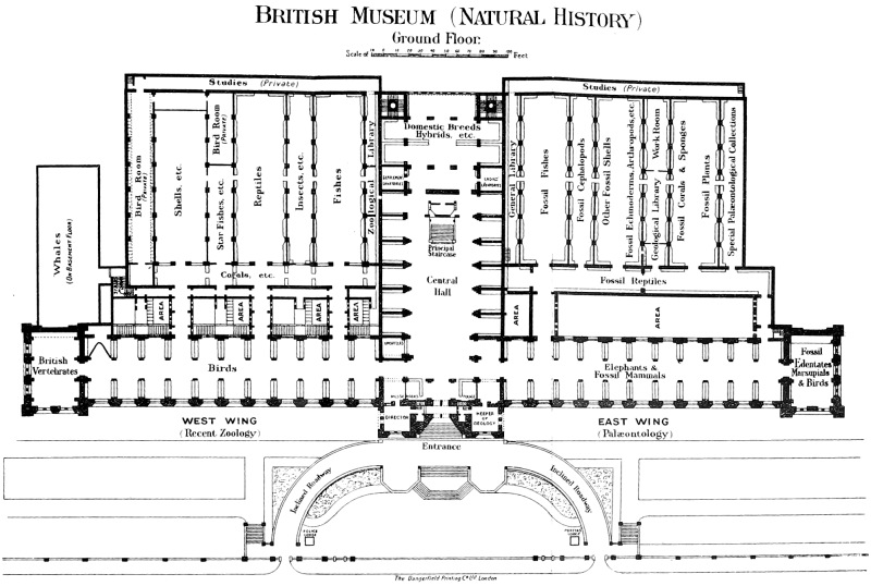
British Museum (Natural History)
Ground Floor.
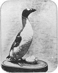
Fig. 15.—The Great Auk or Gare-Fowl (Plautus, or Alca, impennis), and its egg.
Among the multitude of species exhibited in this gallery, which form, however, but a small proportion of the different kinds of Birds known to inhabit the globe, only a few of the more striking can be mentioned. The various types of the Birds-of-Prey are very fully represented: from the Condor of the Andes, the large Sea-Eagle of Bering Strait, and the Great Eagle-Owl of Europe (all of which are placed in separate cases), to the Dwarf Falcon in case 53, which is not much larger than a sparrow, and preys upon insects. Among the large group of Perching-Birds, attention may be directed to the cases of Birds of Paradise and Bower-Birds in the first bay on the left. In separate cases in the sixth bay on the opposite side of the gallery are placed skeletons of the Dodo and Solitaire, large Pigeon-like birds with wings too small for flight, once inhabiting the islands of Mauritius and Rodriguez, respectively, but now extinct. Other cases on the right-hand side of the gallery are occupied by the Game-Birds, and the Wading and Swimming Birds. Here may be noticed a nearly complete series of the genera of Pheasants and Pigeons, showing the various forms. Special attention may be directed to the Great Auk (fig. 15), from the Northern Atlantic, which became extinct only in the last century. Casts of the eggs (fig. 16) of this curious bird are also exhibited. A case in the 7th bay contains a series of Penguins, flightless birds which may be regarded as representing the northern Auks and Guillemots in the southern oceans. Particularly interesting is the great Emperor Penguin, which lays its eggs and rears its young in winter amidst the ice of the Antarctic. Most of the specimens exhibited were obtained during the British Antarctic Expedition of 1839–43, under the command of Captain Sir James Clark Ross.
Other noteworthy types are the Great Bustard, once an inhabitant of England, and the Flamingos; a pair of the latter being exhibited with their nest.
In the first two bays on the right side of the gallery are placed specimens of the Ostrich group, characterised by the flat or raft-like form of the breast-bone. Owing to the rudimentary character of their wings, these Birds lack the power of flight. They include the largest existing Birds, the Ostriches, Emus, and Cassowaries, as well as the small Kiwis (Apteryx) of New Zealand, together with the extinct Moas (Dinornis, etc.), of the same country, and the Roc (Æpyornis) of Madagascar. A fossil egg of the latter is placed alongside eggs of the existing species of the group.
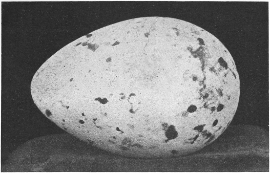
Fig. 16.—Egg of the Great Auk or Gare-Fowl: Size of nature.
Groups of British Birds and Nests.
Down the middle line of the gallery, as well as in many of the bays, are placed groups showing the nesting-habits of various species of British birds. The great value of these groups consists in their absolute truthfulness to nature. The surroundings are not selected by chance or from imagination, but in every case are carefully executed reproductions of those that were present round the individual nest. When it has been possible, the actual rocks, trees, or grass, have been preserved, but in cases where these could not be used, they have been accurately modelled from nature. Great care has also been taken in preserving the natural form and characteristic attitudes of the Birds themselves. Among the more attractive cases are, near the centre of the gallery, a pair of Puffins feeding their single young one, and Black-throated Divers with their eggs in a hollow in the grass on the edge of a mountain-loch in Sutherland. Hen-harriers—the male grey and the female brown—are shown with their nest among the heather from the moorland of the same county. On the left of these is a Peregrine Falcon’s eyrie, on the ledge of a rocky cliff, containing three white downy nestlings. Near by are various species of Ducks, notably the Red-headed Pochard on the sedgy border of a Norfolk mere. In the last bay but one on the right side is a nest of the Heron, in a fir-tree, with the two old birds and three nearly fledged young. Various species of Gulls and a particularly beautiful group of Arctic Terns from the Shetland Islands are exhibited in the middle line towards the west end of the gallery and in the eighth and ninth bays. In the eighth bay on the right side and in the adjoining passage are Plovers, Sandpipers, Snipes, etc., some of which (especially the Ringed and Kentish Plovers) show the wonderful adaptation of the colouring of the eggs and young birds to their natural surroundings for the purpose of concealment. In the second passage leading to the Coral-gallery are Ptarmigan and Capercaillie from Scotland, and in the adjacent part of the middle line Wood-Pigeons and Turtle-Doves building their simple, flat nests of sticks in ivy-clad trees. In the fourth, sixth and seventh bays on the left are Sand-Martins and Kingfishers, showing, by means of sections of the banks of sand or earth, the form and depth of the hole in which the eggs are placed; and also nests of the Swift, Swallow, and House-Martin, all in portions of human habitations.
Pavilion, with British Land and Fresh-water Vertebrates.9
The “pavilion” at the west end of the Bird-gallery is devoted to the exhibition of the land and fresh-water Vertebrated Animals of the British Islands. The larger Mammals and Fishes occupy the wall-case on the north side, which is surmounted with horns. In the two pairs of centre cases is exhibited the series of British Birds, supplemented by the groups, to which reference has been made already. The wall-case on the north side of the archway contains a group of Gannets and other sea-birds from the Bass Rock in the Firth of Forth. On the opposite side are two striking groups with the surroundings true to nature, the one of the Golden Eagle and the other of the Buzzard, both taken in Scotland. Other groups in the pavilion display the Kestrel, the Peregrine Falcon, and the Merlin amid natural surroundings. Among the Mammals, especial attention may be directed to a case of British Hares and Rabbits. In another case may be seen a female Badger and her young; in a third is a group of Otters; in a fourth a vixen Fox with her cubs; in a fifth a Mole-hill with its inhabitants; in a sixth a pair of Martens; in a seventh Polecats and their young; while other cases are devoted to Stoats, Weasels, Hedgehogs, Squirrels, Rats, Mice, etc.
Here it may be mentioned that the animal inhabitants of any country or district are collectively termed its “fauna.” The British Islands in this respect belong to the great zoological region called Palæarctic, or Eastern Holarctic, embracing all Europe, the north of Africa, and the western and northern portions of Asia. As in the case of other islands, the species belonging to groups in which the power of locomotion is limited to land or fresh-water are not numerous compared with those inhabiting large continental tracts. Their numbers can only increase under exceptional circumstances, and have a tendency to diminish as the growth of human population and increase of the area of cultivated land gradually reduce their native haunts. In this way the Brown Bear, the Wolf, the Beaver, and the Wild Boar have disappeared from Britain within the historic period, while other species, such as the Badger, Marten, and Wild Cat, with difficulty maintain a more or less precarious existence. All these were originally derived from the mainland of Europe, probably before the formation of the channel which now separates it from Great Britain. The wider and older channel which separates Ireland from Great Britain has been a greater barrier to the emigration of animal life than that between the latter and the Continent, many species (as the Polecat, Wild Cat, Mole, Squirrel, Dormouse, Harvest-Mouse, Water-Rat, Short-tailed Field-Mouse, Brown Hare, Roedeer, as well as Snakes and Toads) never having crossed what is now the Irish Sea, unless by human agency.
On the other hand, those species that have the power of travelling through the air or traversing the ocean are far less fixed in their habitat; and it results from this that the list of so-called “British Birds” receives accessions from time to time from stragglers which find their way from the European continent or Asia, or even across the Atlantic.
Slight but permanent variations from the continental type may be recognised in many native British species, some of the most marked among vertebrated animals being the Irish Stoat, the Squirrel, the Red Grouse, the St. Kilda Wren, the Coal-Tit, the Goldcrest, and several species of fresh-water fishes, mostly belonging to the genera Salmo and Coregonus. Some of the latter, such as the Vendace, the Gwyniad, and their allies, of which specimens are exhibited in the wall-case in the pavilion, have an extremely local distribution, being found only in certain small groups of mountain lakes.
Of the Seals, only two species are really natives of Britain, the Common Seal (Phoca vitulina) and the great Grey Seal (Halichœrus grypus); specimens of both these are shown in the pavilion.
Those desirous of studying more minutely the characteristics of British Mammals should examine the series of skins and skulls exhibited in a special case on the right side of the central west window.
Coral Gallery.10
Parallel with the Bird-gallery, on the north side (right on entering), and approached by several passages, is a long narrow gallery containing the collection of Corals and Sponges and allied types. Commencing at the eastern end, some of the lowest forms of animal life are exhibited in the wall-case and table-cases; they belong to a group called Protozoa, and, for the greater part, are so minute, that they can be studied only with the microscope; their structure is therefore illustrated chiefly by means of models and figures. The next divisions of the gallery are occupied by the Sponges, most conspicuous among these being a series showing the variations of the common Bath-Sponge (cases 1 and 2), the beautiful flinty Venus’ Flower-basket or Euplectella (fig. 17), the Japanese Glass-rope Sponge or Hyalonema (case 3), and the gigantic Neptune’s Cup or Poterion, of which several specimens are placed on separate stands. Special interest attaches to the case showing the different kinds of Sponges used in commerce.
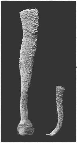
Fig. 17.—Venus’ Flower-Basket (Euplectella imperialis and E. aspergillum). (One-sixth natural size.)
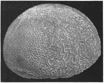
Fig. 18.—Brain-Coral. (Meandrina cerebriformis).
Nearly the whole of the remainder of the gallery is given up to Corals. In life these organisms display an immense variety of form and colour, sometimes presenting a marvellous resemblance to vegetable growths; but the part exhibited in the gallery is merely the dried, hard, horny, or stony basis or supporting skeleton, either of isolated individuals, or of colonies. Corals are allied to the well-known Sea-anemones of the British and other coasts; the combined skeletons of myriads of these animals form the coral-reefs which constitute the bases of thousands of islands in the Indo-Pacific Ocean. Among the larger reef-making species are the Brain-Corals (Meandrina), one of which is shown in figure 18. Near the west end of the gallery is placed a magnificent specimen of the Black Coral of the Mediterranean (Gerardia savalia), obtained off the coast of the island of Eubœa in the Ægean Sea. The drawing in the case shows a magnified view of the “animals” or polyps of this species as they appear in life. In case 13 are specimens and drawings of the Red Coral (Corallium rubrum), so largely used for ornamental purposes, and also of the crimson Organ-pipe Coral (Tubipora musica). Arranged on shelves on the south wall of the western end of this gallery is a series of Pennatulidæ (Sea-pens, Sea-rushes, or Sea-ropes) preserved in spirit. These Zoophytes live at the bottom of the sea, with their lower ends fixed in the sand and mud; the skeleton being never more than a straight internal rod in addition to innumerable microscopic spicules.
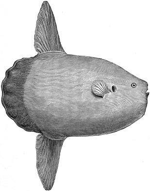
Fig. 19.—Rough Sunfish (Orthagoriscus mola).
Approached through the Coral-gallery, and running backwards at right angles with it, are several galleries containing other portions of the zoological collections.
Fish Gallery.11
I. The Fish-gallery is nearest to the central hall, and contains the exhibited portion of the collection of Fishes. The greater number of specimens, preserved in spirit, are, however, placed for safety in a detached building outside the Museum, where they are available for study under special regulations. The gallery contains mounted examples, models, and skeletons of many of the more remarkable members of the class.
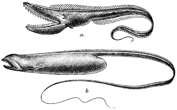
Figs. 20 & 21.—Two Deep-Sea Fishes (a. Gastrostomus bairdi and b. Saccopharynx flagellum).
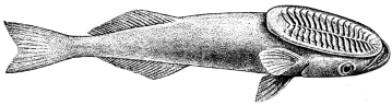
Fig. 22.—Sucker-Fish (Echeneis remora).
The wall-cases on the east side of the gallery (right on entering) contain the fishes with completely bony skeletons (Teleostei); to which division belong by far the greater part of the species now inhabiting the waters of the globe. Large and remarkable examples are placed in separate cases opposite to the wall-cases. As the colours of fishes are very fugitive, and disappear more or less completely after death, most of the mounted examples have been painted. The fishes allied to the Perch, Gurnard, Mackerel, Sword-Fish, Wrasse, Cod, Plaice, Catfish, Salmon, Pike, and Eel are represented by numerous examples. Specially noticeable, so far as size and external form are concerned, are the Sunfishes, Orthagoriscus (fig. 19).
Even more strange are several of the species of deep-sea Fishes exhibited in a table-case in the Fish-gallery, which live at depths where the sun’s rays cannot penetrate, and many of which are self-luminous. Two deep-sea Fishes are shown in the accompanying illustrations (figs. 20 and 21). From another point of view, special attention may be given to the Sucker-Fish or Remora (Echeneis remora), fig. 22, which attaches itself by the sucker on the top of its head to the bodies of Fishes or Turtles, or to the bottom of ships. As it attaches itself back-downwards, the under-parts are coloured dark while the back is light; a condition just the reverse of that obtaining in ordinary Fishes.
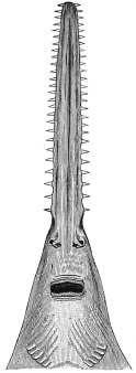
Fig. 23.—Lower View of the Head of a Saw-Fish. (Pristis antiquorum.)
The western or left side of the gallery is devoted to the exhibition of certain very different types of Fishes, which were much more numerously represented in ancient times than at present. The majority have a cartilaginous skeleton. Among these may be specially mentioned the Bichir of the tropical African rivers, the Gar-Pike of North America, the Sturgeons, the Lung-Fishes (Dipnoi) of South America, Africa, and Australia, the Chimæras, and finally the Sharks and Rays. Among the two latter are included the singular Hammer-headed Shark (Zygœna), and the Saw-Fishes (Pristis), which have long projecting flattened snouts, with a row of teeth arranged something like those of a saw on each side (fig. 23). A remarkably large specimen from the coast of British Guiana of a species of this group (Pristis perrotteti) is exhibited. Another very small division of Fishes comprises the Lampreys and Hags, of which a few specimens are shown.
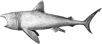
Fig. 24.—The Basking Shark (Selache maxima).
The largest exhibited specimen of the class is the head of a full-grown example of the great Basking Shark (Selache, or Cetorhinus, maxima), fig. 24, captured on the 2nd of March, 1875, near Shanklin, in the Isle of Wight. The length of the entire specimen was twenty-eight feet, but, as the minute size of the teeth indicates, it is a comparatively harmless fish. A smaller female specimen is suspended from the roof; and below this is placed a model of the skeleton of the same species. Near by is a young specimen of another basking species, the Elephant-Shark (Rhinodon typicus), which when adult is said to attain a length of at least fifty feet. It inhabits the Indian and Pacific Oceans.
Models of some of the more remarkable types of extinct Fishes are exhibited in cases on the same side of the gallery.
Insect Gallery.12
II. A small gallery is devoted to the group of Arthropoda or Invertebrate animals with jointed limbs, such as Lobsters, Crabs, Spiders, Centipedes, and Insects.
At the south end of this gallery are exhibited specimens of Crabs and Lobsters. Among the former, special attention may be directed to the specimens of the Giant Crab (Macrocheira) of Japan, and also to the Coconut Crab (Birgus latro), fig. 25, which climbs trees to feed on young cocoanuts, and is related to the Hermit-Crabs. In the central table-cases, besides Crustacea (Crabs, Lobsters, etc.), are displayed representative Scorpions and Spiders, including several examples of the large Bird-eating Spiders of South America.
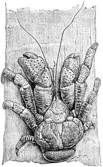
Fig. 25.—The Coconut Crab (Birgus latro).
The northern half of this gallery is devoted to the exhibited series of Insects. Next to the door on the east side are the nests of White Ants or Termites, while on the opposite side are some remarkable cocoons of social Caterpillars of various Moths, and also a series of preparations showing the metamorphoses or changes undergone by Insects as they grow to maturity. At the sides of the gallery are models of various galls, with drawings of the insects which cause them; and others showing the life-histories of certain Beetles, Bees, and Moths. Beneath a shelf, on the west side, are four cabinets containing a collection of British Butterflies and Moths with their caterpillars, prepared and presented by Lord Walsingham; and near by stands a cabinet containing a collection of the Butterflies and Moths of the British islands formed by the late Mr. William Buckler, most of the specimens in which were bred by him during the preparation of his “Larvæ of British Butterflies and Moths.” Above these are maps illustrating the geographical distribution of certain Beetles (Calosoma, Carabus, Julodis and Stigmodera); actual specimens of the insects being placed in position on the maps. The wall-cases at the end of the gallery are devoted exclusively to the nests of Ants, Wasps, and Bees. On the east wall is a large case containing specimens and drawings explaining the structure of Insects. On the west side, next the Walsingham collection, are cabinets containing a selection of British Insects. Lower down are other cabinets in the series of foreign Butterflies; while foreign Moths and other Insects are arranged in adjacent cabinets. In another part of the gallery are exhibited coloured drawings of a few of the smallest Insects known, namely the Mymaridæ, a group of minute parasitic Hymenoptera; and above is a drawing of a House-Fly, enlarged in the same proportion—thirty diameters—to show the contrast. A few specimens of the insects themselves are placed in the microscope below.
In the table-cases in the middle of the gallery are specimens and illustrations of some of the principal families of Insects, with explanations of the characters by which these may be recognised. The first case (next to the west door) contains an introductory series, and then follow the various orders in sequence commencing with the most primitive forms or Aptera, among which Campodea, a small British insect, may be specially noticed.
The classification of Butterflies, Bees and Ants, and Beetles is shown in the last three cases; and attention may be specially directed to a series of drawings illustrating the transformations of Fleas, Gnats, Midges, etc.
The main collection of insects is kept in cabinets in the “Insect Room” in the basement, but is open to students under the regulations mentioned at the end of this guide.
Reptile Gallery.13
III. The long Reptile-gallery contains mounted specimens and skeletons of Reptiles, including Crocodiles, Lizards, Snakes, and Tortoises, as well as restorations or casts of the remains of many groups now entirely extinct. The most noticeable specimen in this gallery is the model of the skeleton of the gigantic extinct North American land Reptile known as Diplodocus carnegii, which measures over eighty feet in length, and was presented by Mr. Andrew Carnegie in 1905. Restored models of the skeletons of the Iguanodon, a British Dinosaur, and of the North American Horned Dinosaur (Triceratops) are also noteworthy.
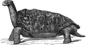
Fig. 26.—Saddle-backed Tortoise (Testudo abingdoni), of Abingdon Island, Galapagos Group.
The Crocodiles and their extinct relatives occupy the cases on the left of the entrance from the Bird-gallery and also a stand in the middle of the gallery; and among these may be specially noticed the Indian Gharial, of which both the skeleton and skin are exhibited. The two small cases on each side of the west doorway are occupied by extinct forms and the peculiar Tuatera Lizard of New Zealand. At the south end of the east side are arranged the Turtles and Tortoises, including examples of the Giant Tortoises of the Galapagos (fig. 26) and Mascarene Islands, as well as a large species (Testudo calcarata) from North Africa. Opposite the Turtles and Tortoises are the Snakes, among which two large Pythons, coloured to nature, form attractive exhibits. Extinct groups occupy a small case on each side of the east door. Beyond these come the Lizards, which occupy the cases opposite the Crocodiles. The series of Old World Monitor Lizards and American Iguanas is specially noteworthy; and attention may be likewise directed to the curious worm-like Amphisbænas, of which the majority inhabit Tropical America. Casts of remains of the extinct marine Ichthyosaurs and Plesiosaurs are shown in the small cases on the sides of the doorway in the east wall.
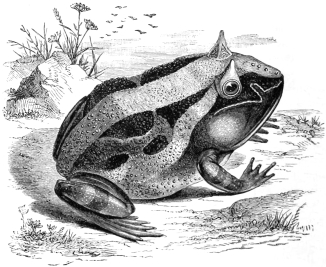
Fig. 27.—The Horned Frog (Ceratophrys cornutus) of Brazil.
Amphibians (Salamanders and Frogs).
In the Reptile-gallery is placed a large table-case containing a selection of the more important forms of Amphibians, which are divided into such as possess a tail:—Salamanders and Newts; and those without tails:—Frogs and Toads. During some period of their existence most of these animals live in the water, when they breathe by gills, and are in this respect akin to Fishes. The comparatively enormous size attained by certain tropical species of Frogs and Toads, such as the South American Horned Frog (fig. 27), should be noticed. The largest representative of the group is the Giant Salamander of Japan and China.
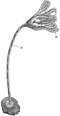
Fig. 28.—A Sea-Lily, the Larval stage of the Rosy Feather-star. (Antedon rosacea), much magnified. a, arms; b, basals; r, radials; s, stalk.
Starfish Gallery.14
IV. A small gallery is devoted to Starfishes and their allies collectively constituting the class Echinoderma. Specimens of these are arranged systematically in table-cases 1–24; and in case 36 are specimens illustrating the anatomy of the skeleton, and models and figures showing the remarkable changes undergone by these animals in the course of their development. In a separate case on the east side of the gallery is a case containing specimens of a large Starfish to illustrate the variability in the number of rays from 4 to 7. In a case on the opposite side is shown Luidia savignei from Mauritius, one of the largest of Starfishes. The Feather-stars (Antedon) are also members of this group; but the most beautiful and remarkable specimens in the gallery are the Sea-Lilies, or Crinoids, collected by the “Challenger” Expedition. One specimen was found attached to an old telegraph-wire taken up in the Caribbean Sea. These deep-sea Crinoids, of which representatives, now extinct, were abundant in earlier periods of the world’s history, are exhibited on tables in the corners of the gallery, by cases 37 and 38. Some of the larval stages of the Feather-stars (fig. 28) resemble stalked Crinoids.
The wall-cases contain representatives of the groups collectively known as Worms. Case 1 contains the Tape-Worms or Cestoda, and the Flukes or Trematoda, the life-history of a species of each being illustrated by specimens, figures, and models. In case 2 the Round-Worms are illustrated by models of Trichina, and the anatomical structure of various other kinds is shown by the aid of diagrams. Case 3 contains specimens of free-living terrestrial and marine Worms, Leeches, and Gephyreans. Case 4 is devoted to specimens of Echinoderms preserved in spirit, especially Holothurians, such as Trepangs or Sea-Cucumbers, the Bêche-de-Mer of the French.
Shell Gallery.15
V. A large gallery is devoted to the great group of Shell-fish or Mollusca, the exhibition of which is, however, mainly restricted to their shells. In some cases the form of the soft parts is, however, shown either by specimens in spirit or by means of models.
In wall-cases on the west side of the gallery is a series of shells arranged on tablets to show the leading structural types, such as univalve, bivalve, multivalve, etc.; then the nature of the outer coat, or “skin”; and, thirdly, some of the more striking styles of ornamentation and colouring. Following these, a division is devoted to the display of the general form of the shells of bivalves, special attention being directed to the nature of the hinge by which the two valves are joined. Fresh-water Mussels (Unionidæ) are selected as examples of great variability in the form of the shell in closely allied species. Near by is a small series of the shells of boring bivalves, many in the various substances they perforate. Alongside are shown in a similar manner modifications in form and structure presented by Univalve or Gastropod shells; many of the shells having been cut to show their internal structure. Specimens of the horny or shelly plate (operculum) closing the mouth of many Gastropod shells are also exhibited. In one compartment are displayed spirit-preparations of Cephalopod Molluscs, such as Octopus, Cuttle-fish, Squids, Nautilus, Argonaut or Paper-Nautilus, etc. Specimens of the horny beaks possessed by all members of this class are also exhibited; and a Pearly Nautilus (fig. 31), with the shell cut in two in order to show the air-chambers and the comparatively small space occupied by the “animal,” will be found of special interest. From the roof are suspended life-sized models of a Giant Squid (Architenthis) and a Giant Octopus. The Cephalopods were extraordinarily numerous in past ages, and many of the fossil forms are exhibited in the Geological Department.
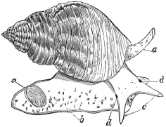
Fig. 29.—The Whelk (Buocinum undatum), a Gastropod Mollusc. a, siphon; b, foot; c, tentacles; d, eyes; e, operculum.
The main shell-collection is contained in four parallel rows of table-cases, the arrangement commencing on the right as the gallery is entered. The first two rows contain the marine forms of Gastropods—a division which includes Snails, Slugs, Whelks (fig. 29), and all those Molluscs which crawl upon the under surface of their bodies; the Cowries, Cones, Volutes, Mitras, and Murexes forming some of the most attractive groups. The two rows of cases on the left contain Land-Shells, Bivalves, and Cephalopods. The Cockles, Oysters, Clams, Piddocks, Teredos, Scallops, and Ark-Shells represent some of the principal types of Bivalves, so called on account of their shells being formed of two pieces or valves. A Giant Clam (fig. 30), at the S. end of the gallery, weighs 310 lbs., and measures 36 inches in length.
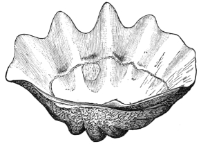
Fig. 30.—Left Valve of the Giant Clam (Tridacna gigantea), a Bivalve Mollusc.
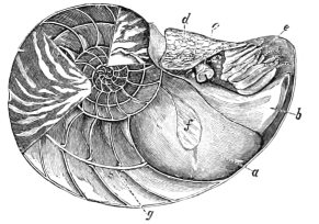
Fig. 31.—The Pearly Nautilus (Nautilus pompilius). a, body; b, funnel; c, eye; d, hood; e, tentacles; f, muscle of attachment to the shell; g, siphuncle.
Sea-Mats, Lamp-shells, and Sea-squirts.
In the same gallery are exhibited examples of three other groups of marine organisms, respectively known as Polyzoa, Brachiopoda, and Tunicata. The Polyzoa live in colonies, and include the so-called Sea-Mat. They are often mistaken for sea-weeds, although in reality they are animals of comparatively high organisation. The Brachiopods have two shells like Bivalve Molluscs, but the valves are dorsal and ventral—that is to say, back and front—instead of right and left. One valve is frequently perforated, hence the name of “Lamp-shells,” from a resemblance to an ancient Roman lamp. Sea-squirts, Tunicates, or Ascidians, are worthy of the attention of the visitor on account of their affinity with the Vertebrate stock, of which they may be regarded as a degenerate type. It is in the larvæ or “Ascidian Tadpoles” that the evidence of Vertebrate relationship is most conspicuous, as shown by certain important characters in the nervous system, skeleton, and respiratory organs.
A series of large shells occupies some of the space in the wall-cases, among which attention may be directed to the thick and often handsomely coloured Helmet-shells (Cassis), which, together with the pink Queen-conch (Strombus), were formerly largely used in the now nearly obsolete art of cameo-cutting. In addition to these, a selection of British shells, and series of the eggs of Molluscs, as well as specimens illustrating the formation of pearls, and other points of special interest connected with Molluscs, are displayed. These specimens include not only the Pearl-oyster, but some of the other shells used in the mother-of-pearl trade.
Whale-Room.
Whale-Room.
Approached by a staircase, leading down from the last (or western-most) of the passages which connect the Bird-gallery with the Coral-gallery, is a separate room in which are placed the specimens of Whales and their relatives. For these, on account of their large size, no other place could be found in the Museum; but the room has, unfortunately, the disadvantage of being too small to display such large animals to full advantage. It is also intersected by columns, which interfere with the complete view of the larger specimens.
As it is almost impracticable to preserve the skins of the larger species of Whales, owing to the oil with which they are saturated, the exhibition of the characters of these animals is carried out by means of their skeletons and artificial models of one side of the body. Complete models, which are much better than actual skins, of many of the smaller kinds, are shown. A general account of the structure and classification of the Cetacea, as Whales are technically termed, with reference to those exhibited in this gallery, will be found in a Special Guide.16
Fresh-water Dolphins.
Narwhal.
On the left side of the entrance is a case containing a stuffed specimen, a skeleton, and several skulls of the Susu, or Fresh-water Dolphin (Platanista gangetica) of the rivers of India, and also of the Dolphins of the Rio de la Plata (Pontoporia blainvillei) and of the River Amazon (Inia geoffroyensis). Among the specimens fronting the visitor as he enters the room, one of the most interesting, on account of its remarkable dentition, is the Narwhal, or Sea-Unicorn. It has only two teeth, which lie horizontally in the upper jaw. In the female both remain permanently concealed within the bone of the jaw, so that this sex is practically toothless; but in the male, while the right tooth remains similarly concealed and rudimentary (as shown in the specimen, by removal of part of the bone which covered it), the left is immensely developed, attaining a length equal to that of half the entire animal, and projecting horizontally from the head in the form of a long, straight, tapering and pointed tusk, spirally grooved on the surface. In rare cases, as in the skull exhibited near the skeleton, both teeth are fully developed, and it is noticeable that in such specimens the direction of the spiral is the same in both tusks.
Sperm-Whale.
To the right of the entrance is placed a specimen of the bony framework of one of the most colossal of animals, the Cachalot, or Sperm-Whale (Physeter macrocephalus), fig. 32, prepared from an old male cast ashore near Thurso, on the north coast of Scotland, in July, 1863, on the estate of Captain D. Macdonald, R.E., by whom it was presented to the Museum. Upon one side of this skeleton has been built the model of the external form of the animal. The Sperm-Whale is the principal source of supply of sperm-oil and spermaceti: the former being obtained by boiling the fat or blubber lying beneath the skin over the whole body. The latter, in a liquid state at the ordinary temperature of the living animal, is contained in cells which fill the immense cavity on the top of the skull. This Whale, which feeds chiefly on Cephalopods (Squids and Cuttle-fishes), but also on Fishes, is distributed throughout the warm and temperate regions of the Atlantic and Pacific Oceans, and sometimes enters British waters.
In order to render this skeleton more instructive, the names have been attached to the principal bones, thus enabling the visitor to trace at a glance the extraordinary modification from the normal mammalian form the huge skull of this species has undergone.
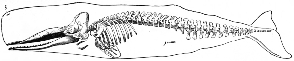
Fig. 32.—An Old Male Sperm-Whale or Cachalot. Skeleton and outline of animal: b, nostril or blowhole; p, rudimentary pelvic bone. Length of specimen 54 feet.
Whalebone Whales.
Most of the largest Cetacea belong to the group called “Whalebone Whales,” in which a series of horny plates termed “whalebone” grow from the palate in place of teeth, and serve to strain the water taken into the mouth from the small marine animals on which these Whales subsist. A representative of this group is the skeleton of the Common Rorqual or Fin-Whale (Balænoptera musculus) in the south-west portion of the room. This Whale, which is sixty-eight feet long, was captured in 1882 in the Moray Firth, Scotland. The flukes of the tail and the back-fin were preserved with the skeleton and are placed above the wall-case behind; the small pelvic bones, and a rudimentary nodule representing the femur or thigh-bone, the only trace of the hind leg of this gigantic animal, are also shown. The external form is modelled in plaster. In front is a skeleton and half-model of the Black or North Atlantic Right-Whale (Balæna glacialis or biscayensis). Below this skeleton is placed a lower jaw of the Greenland Right-Whale (Balæna mysticetus), the species which formerly yielded most of the “whalebone” of commerce, and also a miniature wooden model of the entire animal, on the scale of one inch to the foot.
Remains of extinct Cetaceans—notably the solid bony beaks of the skulls of Beaked Whales (Ziphiidæ) from the Red Crag of the east coast of England—are placed in this gallery. A special table-case, near the Sperm-Whale, shows the curious ear-bones of various Cetaceans, both recent and fossil. These bones are perfectly sufficient for the identification of the kind of Whale from which they were taken. In a case on the opposite side of the gallery is displayed the horny wart, termed by sailors the “bonnet,” found on the nose of the Black Right-Whale.
First Floor.
The upper floors of the wings of the Museum consist merely of single galleries extending along the whole front of the building; for the galleries which run backwards on the ground floor form only a single storey.
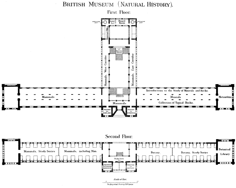
British Museum (Natural History).
First Floor.
Second Floor.
Lower Mammal Gallery.
The Lower Mammal-Gallery is entered from the western corridor of the central hall. Together with the adjacent corridor, it contains the greater part of the exhibited series of recent Mammals, with the exception of the Cetacea, Sirenia, and Proboscidea, which are downstairs, and the orders Primates, Chiroptera, Insectivora, and Rodentia, which are in the upper gallery. As three special guides17 are devoted to these galleries, a very brief notice will serve on this occasion. Both stuffed specimens and skulls and skeletons are exhibited, although the former constitute by far the greater portion of the series. A few remains of extinct types, or plaster reproductions of the same, are introduced here and there; and photographs of living animals are hung on the walls, where will also be found some instructive series showing the modifications assumed by the teeth of certain groups. Wherever possible, the horns and antlers of the Ruminants, as well as the horns of the Rhinoceroses, are placed in juxtaposition to the animals to which they respectively belong.
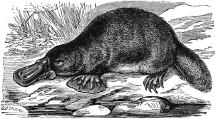
Fig. 33.—The Platypus or Duck-bill (Ornithorhynchus anatinus).
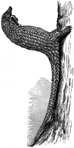
Fig. 34.—The Yellow-bellied Pangolin (Manis tricuspis) in a characteristic attitude.
Ruminants, etc.
The series commences on the right side of the gallery with the lowest forms. Down the middle is a row of large Mammals, comprising various Deer, Seals, and Rhinoceroses. On the right of the entrance a small case contains the Australian Platypus or Duck-bill (fig. 33) and the Echidnas of Australia and New Guinea, which lay eggs, and are the lowest of all living Mammals. In the adjacent bay are the Marsupials, such as Kangaroos, Phalangers, or so-called Opossums, Wombats, and Bandicoots of Australasia, and the true Opossums of America. The eighth and ninth bays on the left side contain the so-called Edentate Mammals, such as the South American Sloths, Ant-eaters, and Armadillos, the Scaly Ant-eaters or Pangolins (fig. 34) of tropical Asia and Africa, and the African Aard-Vark or Ant-Bear. The Sea-Cows, as represented by the Dugong, the Manatee, and the recently extinct Rhytina of Bering Island, are shown in the Geological Department. In the second bay are the Pigs and Hippopotamuses; in the third the Camels, and near by the Chevrotains, or Mouse-Deer. Following these are the Deer (Cervidæ), many of which, as already stated, are placed in the middle line of the gallery. Properly speaking, the Giraffes and their recent and extinct allies, the former represented by the Okapi of Central Africa (fig. 14, p. 41), should come here; but, as already mentioned, it has been found convenient to remove the Giraffe group into the east corridor of the central hall. In one of the cases in the bays stands the Prongbuck or Pronghorn Antelope (fig. 35), the sole living representative of a family characterised by the circumstance that the horns have hollow branched sheaths which are shed annually. Next in order come the Antelopes, a large number of which are placed in the corridors outside the gallery. This series, it may be remarked, is particularly fine, and, in fact, unique. At the last two bays on the right side of the gallery the visitor reaches the Goats; and in the “pavilion,” at the west end of the gallery, he comes to the Sheep, Musk-Oxen, and Oxen, of which there is a magnificent display, both as regards mounted specimens and horns. Many of the cases in the middle of the gallery and the bays have been fitted with artificial groundwork, one of the most striking being the European Reindeer case, for which the materials were brought from Norway.
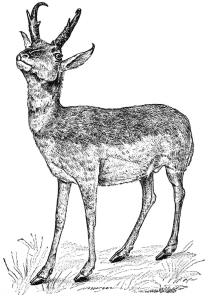
Fig. 35.—Prongbuck or Pronghorn Antelope (Antilocapra americana).
Continuing our survey down the left side of the gallery, the bay next the pavilion and an adjacent case in the middle line contain the Zebras, Wild Asses and the Wild Horse, among which is a specimen of the extinct Quagga. Following this are the Rhinoceroses and Tapirs, some of the former being exhibited in the middle of the gallery. Adult specimens of all the living species except the one-horned Rhinoceros sondaicus of Java are exhibited. The cut (fig. 36) shows the form of the head and the number of the horns in three members of the group.
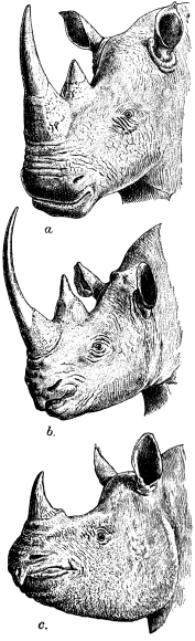
Fig. 36.—Heads of the White Rhinoceros (Rhinoceros simus, a), Black Rhinoceros (R. bicornis, b), and Great Indian Rhinoceros (R. unicornis, c).
Hyraxes.
In a small case by themselves are exhibited the Hyraxes, which represent a subordinal group of Ungulates. In this place should come the Elephants (Proboscidea), but it has been found advisable to exhibit the existing species of this group alongside their extinct relatives in the Geological Department, and in the central hall.
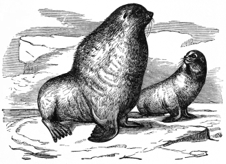
Fig. 37.—Male and Female of the Northern Fur-Seal, or Sea-Bear (Otaria ursina).
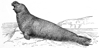
Fig. 38.—Male Elephant-Seal, or Sea-Elephant (Mirounga leonina).
Next in order follow the Seals, Walruses, and Sea-Bears (fig. 37); and after these again, the land Carnivora. Among the former, particular attention may be directed to the specimens of Sea-Elephants or Elephant-Seals (fig. 38) of the Southern Seas and the Pacific coast of California. The visitor should also notice the various smaller southern Seals, obtained during the “Discovery” Expedition, in the case in the bay. Among the land Carnivora, special interest attaches to the huge Brown Bear from Alaska, the black and white Bear-like Great Panda (fig. 39) of North-eastern China, and the case of Tigers, in which both the long-haired Manchurian and the short-coated Indian race are shown.
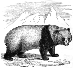
Fig. 39.—Great Panda (Æluropus melanoleucus).
Second Floor.
Upper Mammal Gallery.
The portion open to the public of the gallery on this floor in the western wing of the building contains such of the Mammalian orders (apart from Proboscidea, Cetacea, and Sirenia) as are not shown in the lower gallery. In the first two wall-cases on the right on entering the gallery is displayed a series of Bats, some stuffed, and others in spirit. In the third wall-case are the Insect-eating Mammals (Insectivora), such as Shrew-mice, Moles, Hedgehogs, etc. Next come the Rodents, and then the Lemurs, Monkeys, and Apes, the greater number of the last being exhibited in the large case in the middle of the gallery. Among the more striking specimens may be mentioned the series of Gorillas (fig. 40) and Chimpanzees, and the Proboscis and Snub-nosed Monkeys.
Nearly all the left side of this gallery is devoted to Anthropology, that is to say, to the representation of the zoological characters of the different races of Mankind18; the series including busts, skeletons, skulls, hair, and portraits.
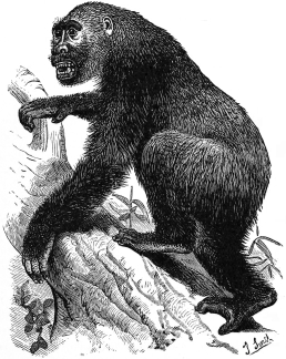
Fig. 40.—The Gorilla (Anthropopithecus gorilla).
At the west end of the portion of this gallery open to the public stands a case exhibiting many of the structural differences distinguishing the man-like Apes from Man himself; and also showing different types of human skulls and the method of measuring the same. On the adjacent screens and partitions are diagrams, photographs, and sketches illustrating hand and finger prints, and identification by means of the latter.
EAST WING.
Ground Floor.
Fossil Collection.
The ground floor of this wing consists, as on the other side of the building, of a gallery running west and east the whole length of the wing in front, of a smaller parallel gallery behind this, and leading from the latter a series of galleries running north and south. With the exception of a certain number of recent skeletons introduced for comparison, and some of the specimens of Elephants and Sirenians or Sea-Cows, the whole of this floor is occupied by the collection of the remains of animals and plants which flourished in geological periods previous to the one in which we are now living. Some of these belong to species still existing upon the earth, but the great majority are extinct. They are arranged mainly upon zoological principles, that is, the groups which are believed to have natural affinities are placed together; but within some of the great divisions thus mapped out, especially of the Invertebrata and Plants, it has been found convenient to adopt a stratigraphical or even geographical grouping, the fossils of different geological formations being kept apart, and those of the British Isles separated from those of foreign localities.
This portion of the Museum is more fully described in the special Guides19 than is possible in the present work.
Elephants, Sea-Cows, and Extinct Mammals.
The front gallery, entered from the central hall, is devoted to Elephants and Sea-Cows, both living and extinct, and to extinct and fossil Mammals of other groups. Down the middle are placed a number of large and striking objects, of too great size to be contained in the wall-cases. The first is a nearly complete skeleton of the American Mastodon (fig. 41), an animal closely allied to modern Elephants, from which it is chiefly distinguished by the characters of its cheek-teeth. This is followed by a skeleton of the existing Indian Elephant (Elephas maximus), and the mounted skin of a tuskless male of the same species, brought home from India by His Majesty King Edward VII., when Prince of Wales. Further down the gallery is the skull of the extinct E. ganesa—remarkable for the immense length of its tusks—from the Siwalik Hills of India; and another of the Mammoth (E. primigenius), with huge curved tusks, in a perfect state of preservation, found in the Brick-earth at Ilford, Essex.20 Then follow skeletons of the great extinct Irish Deer (Cervus giganteus), male and female, the former distinguished by its magnificent spreading antlers, resembling those of a Fallow Deer on a large scale.
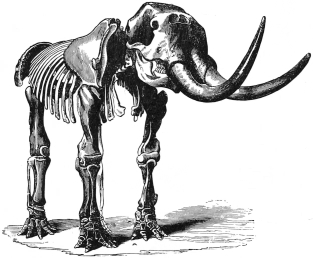
Fig. 41.—Skeleton of the American Mastodon (Mastodon americanus). Greatly reduced.
The next central case is occupied by the skulls and portions of jaws of a remarkable horned hoofed quadruped, Arsinoïtherium (fig. 42), from the Upper Eocene of the Fayum, Egypt. It belongs to an ancient group not closely related to any living animal.
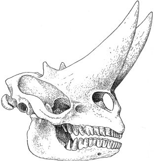
Fig. 42.—Skull of Arsinoïtherium zitteli, from the Upper Eocene Strata of the Fayum, Egypt. About 1/12th natural size.
Equally peculiar is the Toxodon (fig. 43) from the Pampas of S. America, of which a model of an entire skeleton is exhibited.
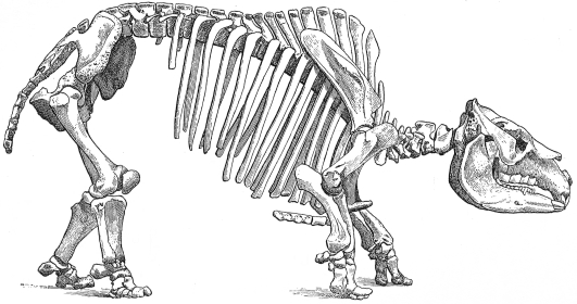
Fig. 43.—Skeleton of the Toxodon (Toxodon platensis).
From the Pampas Formation of Argentina. About 1/18th natural size.
Near by is placed a model of a skeleton of the Dinoceras (fig. 44), one of the most remarkable of the many wonderful forms of animal life discovered in the Tertiary beds of the western portion of the United States of America. This animal combines in some respects the characters of a Rhinoceros with those of an Elephant, and has others altogether special to itself. The group to which it belonged became extinct in the Oligocene, or Middle Tertiary, period, without leaving any successors.
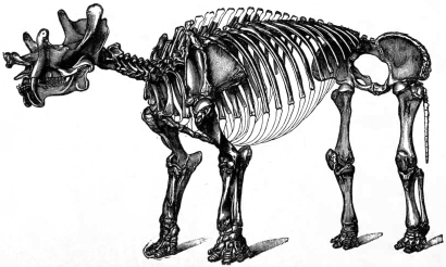
Fig. 44.—Skeleton of Dinoceras. From the Eocene of Wyoming, U.S.A. One-thirtieth natural size.
Sea-Cows.
Near the pavilion is a skeleton (fig. 45) of an interesting animal, the Northern Sea-Cow (Rhytina gigas or stelleri), the last resort of which was Bering Island in the North Pacific, where it was completely exterminated towards the close of the eighteenth century. In the same case is placed the skeleton of a smaller allied animal, the Halitherium, from the Oligocene of South Germany. These, with their existing representatives, the Manatee and Dugong, constitute the order Sirenia, aquatic Mammals of fish-like form, presenting considerable external resemblance to Cetacea (Whales and Dolphins), but differing in many points of structure and habit. All the exhibited specimens of the group, both living and extinct, are shown in this gallery.
Fossil remains of Man.
The wall-cases on the south side (right on entering) contain typical series of chipped and polished flint implements of human workmanship, and also remains of Man found, under circumstances which may justify the appellation of “fossil,” in caves or Pleistocene deposits, associated with the bones of Mammals either completely or locally extinct. Then follow in systematic order the bones and teeth of the other Primates, the Carnivora, Ungulata, and Sirenia.
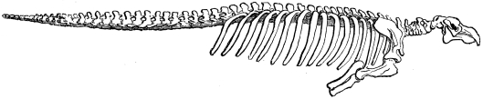
Fig. 45.—Skeleton of the Northern Sea-Cow (Rhytina gigas or stelleri). From Bering Island. Length of specimen, 19 feet 6 inches.
Elephants and Ruminants.
The greater part of the north side of the gallery is devoted to the exhibition of the remains of Proboscidea (Dinotheres, Mastodons, and Elephants), as well as teeth and skulls of the two existing species of Elephants. The forerunners of the Proboscidea from the Eocene strata of Egypt, as represented by Palæomastodon and Mœritherium, are of special interest. Attention may likewise be directed to the skull of the gigantic Ruminant Sivatherium (fig. 46), from the Siwalik deposits of Northern India, an ally of the living African Okapi (p. 41), with which it is connected by Helladotherium of the Grecian Tertiary deposits; another still more nearly allied type being Samotherium, from the Isle of Samos, of which a skull (fig. 47) is exhibited.
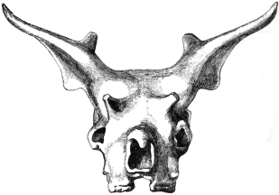
Fig. 46.—Skull of Sivatherium giganteum, an Extinct Ruminant from the Pliocene Deposits of the Siwalik Hills, India. About 1/13th natural size.
Edentates.
In the “pavilion,” or large room at the end of the gallery, are skeletons and bones of the members of the order Edentata, mostly from South America, including fine specimens of the great Ground-Sloths, the largest of which, the Megatherium, is shown in the act of rearing itself on its hind-legs and powerful tail to seize and tear down the branches of a tree in order to feed upon the leaves. That this was the habit of this huge animal is clearly indicated by the structure of its bones and teeth. The mounted specimen is not an actual skeleton, but is composed of plaster-casts of the real bones, most of which are shown in the wall-case at the north side of the room. Of the Mylodon, a smaller but nearly allied animal, an almost perfect skeleton is exhibited in a glass case near the Megatherium. Near by is a portion of the skin of a closely similar animal (Grypotherium listai), from a cave in Patagonia, showing the hair, and also the nodules of bone embedded in the hide.