Volume 4 / Part 2 — CHAPTER VI OPERATIONS INVOLVING THE NASO-PHARYNX: OPERATIONS FOR RETROPHARYNGEAL ABSCESS: OPERATIONS FOR NASO-PHARYNGEAL ADENOIDS
Reading edition
METHODS OF OBTAINING ACCESS TO THE NASO-PHARYNX THROUGH THE NOSE
Many growths in the naso-pharynx, whether originating in the space or descending into it from the posterior choanæ, can be removed by the following method.
Indications. This operation is indicated for the ordinary mucous polypus of the nose when presenting in the post-nasal space. Polypoid masses of the ethmoid may project through the posterior cavity and are removed in the same way. A naso-pharyngeal polypus (also called choanal polypus, post-nasal polypus, or benign pharyngeal polypus) is easily removed by this procedure. Innocent tumours of the post-nasal space, such as papilloma, adenoma, fibroma, and cysts, can be removed by the same method.
Under cocaine. Cocaine and adrenalin (see p. 572) should be carefully applied to the septum and turbinals, as it is the passage of the instrument from the front which is often the most painful part of the proceeding. The pharynx should be lightly sprayed with a 5% solution of cocaine so as to check reflex action.
While the patient is seated in the ordinary examination chair the surgeon stands at his left hand and introduces a looped snare (Fig. 312, p. 613) through the nostril most suitable for approaching the root of the growth. When the snare has reached the post-nasal space, the surgeon introduces the purified forefinger of the left hand through the mouth and up behind the soft palate, as in Fig. 291. Here it serves to manipulate the loop over the growth, and holds it close to the root of the pedicle while the snare is pulled home.
A few minutes should be allowed to elapse to permit the patient to recover from the unpleasant manipulation, and also to allow of coagulation of the strangulated blood-vessels. The growth should not be cut through, as it is wiser to pluck it from its attachment by a quick movement of avulsion.
The growth may come away with the snare through the nostril, or may fall into the pharynx and be expectorated.
In fairly roomy nostrils a stout polypus forceps can be used instead of the snare.
Under chloroform. In nervous subjects the same method should be carried out under a general anæsthetic, care being taken that the growth does not cause embarrassment by occluding the larynx.
Under chloroform, of course, more extensive operations can be carried out on the post-nasal space. The pedicle can be attacked with a pair of scissors with long handles, short blades, and slightly curved on the flat. These are introduced through that nostril which appears to be in most direct line with the pedicle, to act as a raspatory, and then cut through the base of the growth. In some cases an instrument such as Langenbeck’s elevator (Fig. 338) will prove useful if introduced through the nostril. The growth is then removed through the mouth by a twisting movement with a strong volsella.
OPERATIONS FOR OBTAINING ACCESS TO THE NASO-PHARYNX THROUGH THE MOUTH
Many growths in the naso-pharynx can be removed through the mouth, without preliminary operations through the face or through the hard or soft palate.
Indications. The following method of access to the naso-pharynx is chiefly called for in true fibroma of the naso-pharynx, otherwise called naso-pharyngeal polypus, fibroid tumour of the base of the skull, fibroid tumour of the naso-pharynx, retro-maxillary polypus, or juvenile sarcoma of the naso-pharynx.
It is also a plan of procedure which may be called for in any very large, innocent tumours of the naso-pharynx, particularly in cases where nasal stenosis prevents access from the nostrils. It would be a suitable method in any operable cases of malignant disease of the post-nasal space.
Operation. The patient is chloroformed and placed in the position of Rose (hanging head). The mouth being propped open, and the tongue drawn forward, the tumour is first explored with the forefinger, to detect and detach any secondary adhesions. A raspatory which works laterally is next passed from one side of the naso-pharynx to the other above the growth. A rugine which works in a sagittal plane is then introduced below the tumour and made to pass upwards behind it—the reverse movement of Gottstein’s curette in the removal of adenoids (Fig. 350). This movement is facilitated by securely gripping the tumour and dragging it forwards with a stout pair of alligator or volsella forceps. The tumour can thus be so liberated that, with some twisting movements, it can sometimes be extracted entire—often dragging down with it through the naso-pharynx any prolongations thrown forward into the nose.
It is useless to attack such growths as true fibroma of the naso-pharynx with an ordinary wire snare, or such an instrument as a pair of adenoid forceps. For these firm tumours, specially powerful forceps have been designed by Doyen and Escat.
Hæmorrhage is apt to be sudden and copious, but the more rapidly and completely the growth is removed the sooner will bleeding cease—even spontaneously. After complete removal firm pressure with a marine sponge will generally check it. A post-nasal plug should be avoided, and is not usually required. Incomplete operations not only start hæmorrhage but may start septic absorption.
Modifications. (a) Preliminary laryngotomy. A preliminary laryngotomy, strongly recommended by J. W. Bond and extensively adopted by Butlin, adds nothing to the dangers of the case. It allows of the laryngo-pharynx being packed, so that there is no anxiety in regard to the descent of blood into the lungs, and it permits the steady administration of the anæsthetic through the laryngotomy canula. The surgeon is thus relieved of two great anxieties, and can devote himself without embarrassment to more deliberate operation.
The laryngotomy tube can be removed as soon as the patient recovers consciousness and all hæmorrhage has ceased.
(b) Division of the soft palate. In addition to the operation of laryngotomy, the following procedure will allow of more deliberate removal.
The soft palate and uvula are carefully divided in the middle line, and a silk ligature is placed through each lateral half so that they can be held forward out of the way. This gives more direct access to the post-nasal tumour, and if then found to crowd the cavity too closely to allow of manipulation, the posterior part of the hard palate can be chiselled away in the middle line. At the conclusion of the operation the divided palate is carefully united in the middle line (see Vol. II).
Selection of method. In some cases operation through the mouth may have to be combined with a second operation from the front—such as the method of Moure (see p. 619) or that of Rouge (see p. 622).
Rapidity of operation is important, as, once the pedicle has been cut through, or the body of the tumour removed, the hæmorrhage tends to subside spontaneously, or is quickly controlled by packing.
The hanging head (Rose) or the Trendelenburg position is generally recommended.
The preliminary laryngotomy seems desirable in all cases. The division of the palate should be avoided if possible. It may not always unite, and is less likely to do so if subsequent operations are required. The soft palate is very elastic, and in some cases it can be tied out of the way by means of a soft rubber catheter passed along the floor of the nose, and out through the mouth.
Ligature of the external carotid, strongly recommended by Chevalier Jackson87, is not necessary unless the patient is very anæmic or weak from former hæmorrhages. It should then be only a temporary ligature (see Vol. I, p. 383).
Hæmorrhage, as already remarked, is chiefly guarded against by rapid and complete operation. The preliminary use of adrenalin and cocaine, the administration of lactate of calcium, and the other methods recommended for the prevention of bleeding (see p. 574) should be carefully attended to. But in every case preparation should be made beforehand for ligature of the external carotids and for saline infusion.
OPERATION FOR RETROPHARYNGEAL ABSCESS
Indications. The disease is serious, and when not diagnosed almost inevitably ends in death. Before the abscess bursts death may result from spasm of the glottis, laryngeal œdema, or asphyxia. The affection runs its course in 5 to 10 days, and if the abscess opens spontaneously death almost inevitably results—either from suffocation, or septic pneumonia, or cardiac failure.
Operation. When the diagnosis is settled intervention should be prompt. It is not necessary to wait for distinct fluctuation. The pus focus may be so difficult of manipulation in an infant, and the pharyngeal muscle may be so thick and indurated, that it is practically impossible, even in the later stages of retropharyngeal abscess, to detect the presence of pus by palpation.88
The evacuation of the abscess through the mouth was formerly looked upon as dangerous, owing to the difficulty of drainage, the fear of pus burrowing behind the œsophagus, and the risk of flooding the larynx with pus. The more difficult plan of opening it from the neck was generally recommended. The majority of cases can be opened through the mouth with perfect safety.
No general or local anæsthetic is administered, but everything necessary for an immediate tracheotomy should be ready at hand. No gag should be employed, a tongue depressor or the operator’s left forefinger being sufficient both to keep the mouth open and act as a guide. The infant is swaddled in a shawl so as to completely control the movements of the extremities and is then laid on its side on a low pillow, and held by a trustworthy assistant. The sinus-forceps used for opening a peritonsillar abscess are thrust into the most prominent part of the swelling, and the opening enlarged by separating the blades as they are withdrawn. A slender sharp-pointed bistoury, guarded and guided by the index-finger, may be used instead of the forceps. The pus will pour out through the nose and mouth. The incision of the pharynx should be free, deep and long, and directed against the posterior wall of the pharynx and as close to the median line as possible, so as to avoid any chance of wounding the internal carotid.
The surgeon may feel more security if, with the same precautions and with the patient in the same position, he first aspirates the pus cavity.
If more accustomed to it, he may also prefer to have the child flat on its back, with the head overhanging the edge of the table.
Suffocation may be so imminent when the patient is first seen that a preliminary tracheotomy is required.
The external operation, which leaves a certain scar, is reserved for some rare cases—as when the abscess is too low to be easily reached through the mouth, when the spasm of the masseters cannot be overcome, when a large pulsating vessel is noticed in front of the abscess, and when the abscess points towards the neck. It is also the suitable one for the chronic and generally tubercular form of abscess more commonly met with in older patients.89
The external operation is made through an incision along the posterior border of the sterno-mastoid muscle, and the dissection is carried behind the large vessels of the neck and in front of the prevertebral muscles.
After-treatment. The after-care of the patient will require consideration, since the disease is generally met with in the feeble and ill nourished.
If the abscess be opened in good time the patient is at once relieved and begins to recover rapidly.
REMOVAL OF NASO-PHARYNGEAL ADENOIDS
Indications. The removal of naso-pharyngeal adenoids is not called for simply because they are accidentally discovered to be present, nor does the need of operation depend solely on the size of the growths or the nasal obstruction they produce. Adenoids require removal whenever the symptoms attributable to them call for relief. These symptoms may be arranged in three groups, according as they are those (i) of nasal stenosis, (ii) of secondary septic infection, or (iii) of reflex effects.
(i) Amongst the first are mouth-breathing and all the numerous sequelæ, including facial, buccal, dental, and thoracic deformities. It must not be forgotten that mouth-breathing may never be present, and yet deformities of the chest or septic or reflex results can be produced by a small amount of growth in the post-nasal space.
(ii) Amongst secondary septic infections are catarrhal conditions of the Eustachian tube and otitis media, and catarrhal infection of any part of the air-passages. Cervical glands and so-called ‘glandular fever’ occur in this group, as do septic gastritis and other conditions caused by the conveyance of sepsis to more distant parts.
(iii) Various reflex effects are sometimes attributable to naso-pharyngeal adenoids. Laryngismus stridulus, reflex cough, chorea, convulsions, night-terrors, enuresis nocturna, and aprosexia are some of the ailments which may justify operation on Luschka’s tonsil.
As it is chiefly in children that this operation is required it is important to see that they are free from indication of infectious fevers. The operation should be postponed until any acute catarrh has subsided. If there be otorrhœa the ears should receive suitable cleansing treatment for a week or two beforehand. The condition of the teeth requires attention.
The operation is so frequently carried out in private houses that it is well to make inquiries into the health of the members of the household, recent illness, and sanitation. When possible, a large, airy room with a south aspect should be chosen.
Operation. In adults it is possible to carry out the operation under cocaine. On the Continent, particularly in hospital practice, it is often done without any anæsthetic at all. In this country general anæsthesia is almost the universal custom. Opinion is divided as to which is the safest and most suitable anæsthetic to employ.
When the removal of tonsils or other operation is not carried out at the same time, an anæsthesia of less than a minute is sufficient. In adults, and in children over 10 years of age, nitrous oxide does excellently. Younger children are apt to be alarmed by the face-piece and apparatus necessary for nitrous oxide, and this gas does not seem so suitable for them as for adults. In younger children chloride of ethyl is extensively employed on the Continent, but has not met with general favour here.
When the tonsils require removal, or any other operation on the upper air-passages is carried out at the same time, and in young children generally, an anæsthesia allowing of more deliberation is desirable. For this, some operators employ ether,—preceded or not by nitrous oxide. But the well-known objections to pure ether in the surgery of the air-passages have caused the preference to be given to chloroform, or to one of the mixtures of chloroform and ether.
The patient should lie quite flat on the operating table, with only a low pillow or folded towel under the head. The anæsthetist, who takes charge of the gag and flexes or rotates the head as directed, stands at the end of the table. At the patient’s right hand stands the surgeon, and within easy reach are his instruments, sponges, and iced water. Standing on the same side and behind him is the nurse. Her duty is to soothe the patient while passing into unconsciousness, and later on to roll him well over on to his right side as the operation finishes.
The operation can be carried out more correctly, rapidly, safely, and comfortably if the surgeon be armed with an electric forehead search-light (see p. 571). Failing this, the table should be brought close up and parallel to a window, with the patient’s right hand next the light.
Surgeons differ as to the degree of anæsthesia desirable. Some like it to be quite light, so that the patient is all the time in the struggling stage and requires his hands to be controlled by the nurse. I think this is quite as dangerous as when the anæsthesia is pushed until the patient is relaxed, with the corneal reflex just abolished, and the swallowing and coughing reflexes still present.
Fig. 349. Adenoid Curette. StClair Thomson’s modification.

When the anæsthetic is administered steadily, with plenty of air, a degree of unconsciousness is generally secured which will allow of an operation lasting two or three minutes without any further adminis[t]ration. Should the patient show signs of recovering consciousness more chloroform can be given from a Junker’s apparatus.
Fig. 350. The Removal of Naso-pharyngeal Adenoids. Semi-diagrammatic illustration to show how the curette revolves around an axis which moves from a, through b and c, to d. The growth is pressed into the fenestra of the instrument in the a' position, and when the sweeping movement has brought it to d', it is detached and caught in the cage.
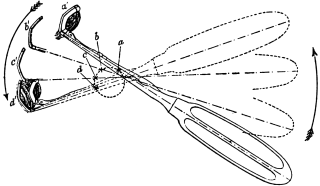
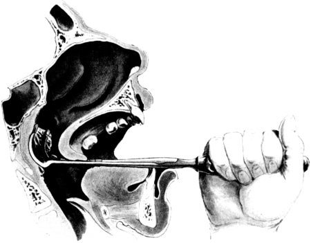
The anæsthetist then opens the mouth with a suitable gag, such as Doyen’s or Mason’s, and maintains the patient’s head exactly in the middle line of the body. Directing the electric search-light into the pharynx, the surgeon depresses the tongue with a spatula in the left hand, while with the right he holds the adenoid curette—some modification of the original Gottstein model (Fig. 349). This is best seized firmly dagger-wise (Fig. 351). It is then introduced along the tongue and slipped up into the post-nasal space. Once safely behind the soft palate and kept straight in the middle line, no harm can be done. Dropping the tongue depressor, the surgeon depresses the handle of his instrument until the beak of it is felt in contact with the posterior free margin of the septum. Pressing the cutting blade firmly and steadily along this it is swept upwards, backwards, and downwards along the vault of the naso-pharynx, while the curette revolves around an imaginary centre in its shaft (Fig. 350). As the instrument is withdrawn from the pharynx, its cage will be found to contain the adenoid growth, removed en bloc and generally complete (Fig. 351). Should the growth slip from the cage, or remain semi-detached from the posterior pharyngeal wall, it can be seized and lifted from the throat with a pair of post-nasal forceps (Fig. 287).
Fig. 351. Removal of Naso-pharyngeal Adenoids. The growth is shown as partially removed from its attachment, and bulging into the cage of the instrument which opens to receive it.
The rush of blood which now takes place is met by rolling the patient well over to his right side, with his face over the edge of the table, so that the blood can run into the right cheek and so out through the mouth. With the patient on his side there is no anxiety of asphyxia from descent of blood or fragments of growth into the trachea, and the surgeon can more deliberately explore the post-nasal space and, with a simple adenoid curette, remove any lateral remains of growth which may have escaped the caged curette.
Sponges are merely used to cleanse the mouth and pharynx in order to make sure that no semi-detached fragments are left behind. If present, tonsils can be conveniently removed at this stage.
Bleeding, which may be very free for a minute or two without any cause for anxiety, is promptly arrested by freely sluicing the patient’s face and neck with ice-cold water.
After-treatment. The patient is put back to bed, lying well over to one side. He should not be allowed to lie on his back, or left unattended, until consciousness has returned. Collapse may occur at this time, generally as a precursor of vomiting, or blood may be vomited and then, owing to the patient’s semi-conscious condition, may be drawn into the trachea.
Ice may be sucked. After a few hours, if there be no vomiting, barley-water, lemonade, tea, thin beef-tea, or beef jelly can be given. Milk and milky food should be avoided. An aperient should be given the same evening, as any foul breath or feverish condition is more likely to be due to blood and mucus in the stomach than to local sepsis. The mouth is kept cleansed with the tooth-brush and an alkaline wash.
It is best to avoid local treatment for the nose. At the end of a few hours the patient is encouraged to clean the nose, and if he be supplied with abundance of fresh air through freely opened windows, the wound in the post-nasal space will heal promptly without any local or general reaction. Occasionally an alkaline nose lotion is required if there has been much secondary rhinitis, or if the child be kept in vitiated air.
One day in bed is generally sufficient, and a child may be allowed out in two or three days, though fatigue should be avoided for a week. Suitable after-treatment in the way of breathing exercises, gymnastics, speech correction, and tonics is often needed. Relief of nasal stenosis may require completion by attention to the condition of the turbinals and septum.
The operation in adults is performed under nitrous oxide. This can be carried out in exactly the same way as that already described, but some surgeons prefer to have the patient sitting up in a dentist’s chair. In that case, after the removal of the mass of growth, the patient’s head is thrown forward between his knees.
Difficulties and dangers. It may be said that the operation itself, carried out with usual care and in a patient who is not a hæmophilic, is free from danger. The chief anxiety is from the anæsthetic, and no inconsiderable number of deaths from this cause have been reported. When possible, it is well to secure the services of an expert anæsthetist who is well used to laryngological work, and accustomed to the operator’s particular methods.
Hæmorrhage may be brisk, even profuse, for a few minutes, but as a rule it promptly ceases if the operation be completed, the patient well rolled to one side, the air thoroughfare left clear so as to allow free breathing and avoid congestion, and the gag removed to permit swallowing and diminish pharyngeal reflexes. The more rapidly and completely the operation is executed, the less will be the bleeding. It not infrequently originates from semi-detached fragments of growth. Even when the hæmorrhage is profuse it is better to push on and complete the removal of growth before attempting to check it. The value of free applications of ice-cold water cannot be exaggerated (see p. 574). In many cases bleeding is maintained by the surgeon’s anxious efforts to stop it with sponging, pressure, or the application of styptics. The greatest danger arises in the case of hæmophilics. If this diathesis be undoubtedly present, the operation should be avoided. If only suspected, more care than usual should be taken in preparing the patient for operation, and lactate of calcium in 15 to 30 grain doses twice a day might be given for two or three days beforehand.
When bleeding persists it is met by keeping the patient very quiet and free from alarm, in a cool and well-ventilated room, and only lightly covered with clothes. Ice is given to suck and applied on each side of the neck, while iced cloths are applied to the face and forehead. Clots are blown out of the nose so as to permit the access of fresh cold air to the post-nasal space. With a pipette, or a pledget of cotton-wool, a few drops of adrenalin can be trickled into the nostril and allowed to run backward. If these measures fail—as they rarely do—the post-nasal space must be plugged (see p. 575). When hæmorrhage takes place after the removal of adenoids and tonsils, it will generally be found that the source of it is in the tonsillar area.
The uvula may retract strongly at the moment of introducing the curette and then get crushed against the posterior pharyngeal wall: or it may be seized by mistake with the post-nasal forceps and be torn away. The same instrument has sometimes been responsible for fracturing the posterior margin of the septum, injuring the Eustachian cushion, and tearing off strips of mucosa from the pharynx. These complications are avoided by using a frontal search-light, operating deliberately, and abandoning the forceps in favour of the curette. This latter instrument can be manipulated without these risks if it be first guided safely behind the uvula and then used more like a carpenter’s adze than a curette. The stroke with the caged curette should be carried through in one movement and exactly in the middle line of the body, but always on the posterior wall. There is no need to attempt removal of adenoid tissue on the lateral walls. This atrophies if the main mass is removed, and the fossa of Rosenmüller can be cleared out with the forefinger.
Local sepsis rarely follows if the precautions described be observed, and local douching is avoided. Any local fœtor—if not arising from the stomach—is generally traceable to some semi-detached fragment which can be removed from the posterior wall with a wire snare (Fig. 312) or a pair of forceps (Fig. 287).
Deafness, earache, and otitis media will sometimes follow the operation, even when the use of a nasal douche has been carefully avoided. They are best met by warm applications, disinfection of the ear with carbolic lotion (5%), and early incision of the drum under nitrous oxide gas.
Other methods of operation. Removal through the nasal chambers—the route originally used by Meyer for his ring-knife—is not to be recommended.
Treatment of the growth with the galvano-cautery, introduced through the mouth, is difficult, risky, and unsatisfactory.
The use of Loewenberg’s forceps, or some modification (Fig. 287), is generally abandoned by any one who has become accustomed to the Gottstein’s curette. A small pair of forceps is, however, very serviceable in quite young children in whom the post-nasal space may be so small as to prevent the manœuvring of any form of curette.
The position with the extended head over the end of the table—Rose’s position—increases the congestion and hæmorrhage, and by throwing forward the cervical vertebræ makes the approach to the roof of the naso-pharynx more difficult.