Description of Plates
Reading edition
Plate I.
Fig. 1.
Diagrammatic sketches showing the development of a spermatozoon from a spermatogonium.
Fig. 2.
Diagrammatic sketch showing the minute structure of a spermatozoon. The middle piece is made comparatively thick in order to bring out the finer structures. Adapted from Ellenberger.
Plate II.
Fig. 3.
Testicle of bull, showing extensive degeneration and necrosis.
Plate III.
Fig. 4.
Inner part of wall of ductus deferens. Normal. × 230.
Fig. 5.
Same, but showing extensive degeneration and exfoliation of the lining membrane. × 230.
Plate IV.
Fig. 6.
Ductus deferens, showing entire exfoliation of the lining membrane. The lumen is filled with a cellular debris. × 50. 61
Fig. 7.
Same, showing the degeneration of the lining membrane, and debris in lumen. × 230.
Plate V.
Fig. 8.
Seminal vesicle of bull. High power section showing the normal structure of the vesicular cavities.
Fig. 9.
Seminal vesicle of bull. Low power. The membrane lining the cavities is degenerated and exfoliated. The cavities are filled with cellular debris, and exudates. There is some increase in the interstitial tissue, and atrophy of some of vesicular cavities.
Plate VI.
Fig. 10.
Low power section of seminal vesicle of bull. The condition is about the same as in Fig. 9 except that it is not quite as severe.
Fig. 11.
Same as Fig. 10. High power.
Plate VII.
Fig. 12.
Testicular tubule showing normal spermatogenesis. × 230.
Fig. 13.
Testicular tubule showing no evidence of mitosis. × 230.
Fig. 14.
Testicular tubule. The spermatogenic epithelium is beginning to degenerate and become cast off into the lumen. × 230.
Fig. 15.
Testicular tubule, showing almost total exfoliation of the spermatogenic epithelium. × 230.
Plate VIII.
Fig. 16.
Testicular tubule. The seminal epithelium is entirely degenerated. The membrana propria is markedly thickened. × 230.
Fig. 17.
Testicular tubule. The tubule is undergoing atrophy and degeneration. The interstitial connective tissue is much increased in amount. × 230.
Fig. 18.
Same as Fig. 17, except that it is in the more advanced stages.
Fig. 19.
Testicular tubule. The spermatogenic epithelium has undergone a sort of hydropic degeneration. The interstitial connective tissue has become much increased in amount and has undergone cellular infiltration.
Plate IX.
Fig. 20.
Normal structure of epididymis tubule. × 140.
Fig. 21.
Epididymis tubule showing exfoliation of the lining membrane, and cellular debris in the lumen. × 140.
Fig. 22.
Atrophy and degeneration of epididymis tubule. The interstitial connective tissue is much increased in amount. × 140.
Fig. 23.
Same, but in more advanced stage. × 140.
Plate X.
Fig. 24.
About the same as Fig. 22. × 140.
Fig. 25.
Marked degeneration of epididymis tubule. There is a cellular infiltration of the interstitial tissue. × 60.
Plate XI.
Fig. 26.
Spermatozoon, showing constriction at middle of head. The head is also somewhat contorted. × 670.
Fig. 27.
Spermatozoon. Pear shaped head. × 670.
Fig. 28.
Spermatozoon. The head is quite long, and pointed at its posterior end. × 670.
Fig. 29.
Spermatozoon, showing a constriction at middle of the head. × 670.
Plate XII.
Fig. 30.
Microcephalic spermatozoon. × 670.
Fig. 31.
Spermatozoon. The head is small, and pear shaped. × 670.
Fig. 32.
Macrocephalic sperm. The middle piece is much thickened. × 670.
Fig. 33.
Tailless spermatozoa. × 670.
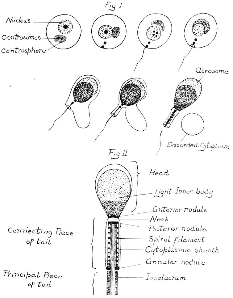
PLATE I
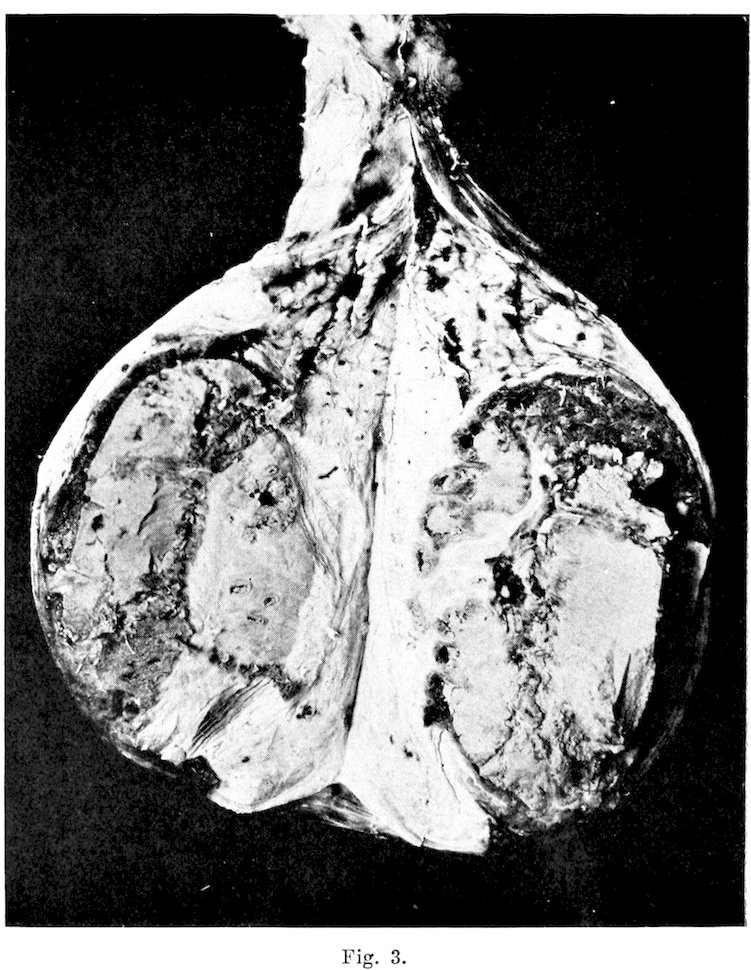
PLATE II
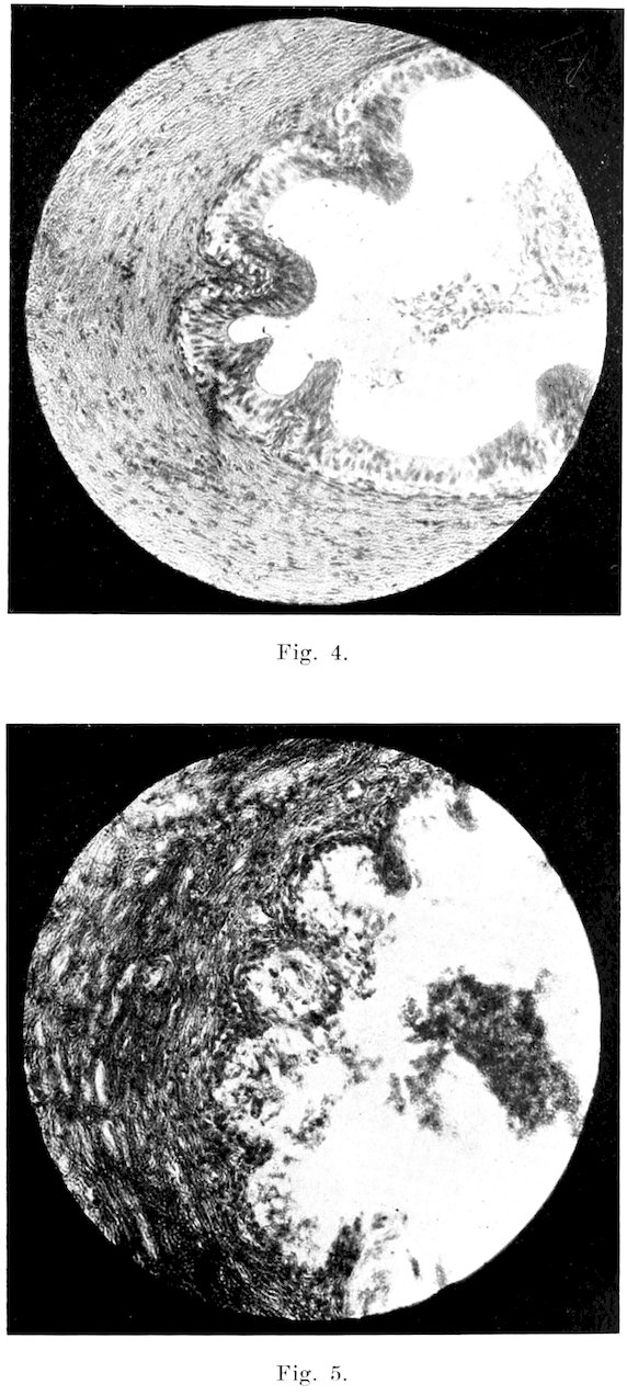
PLATE III
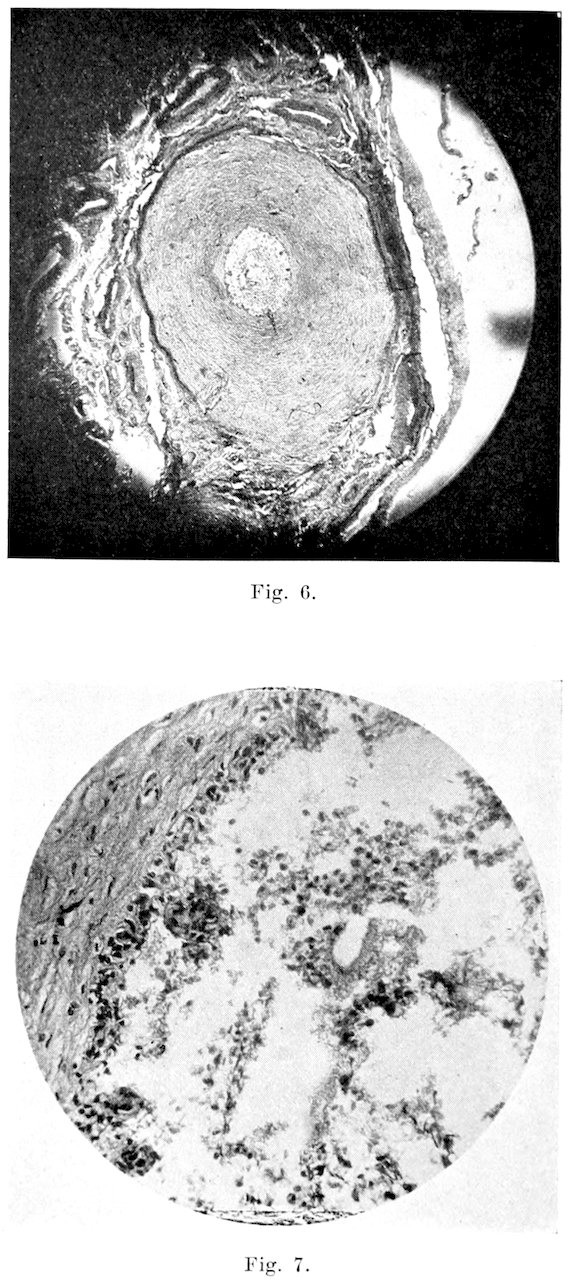
PLATE IV
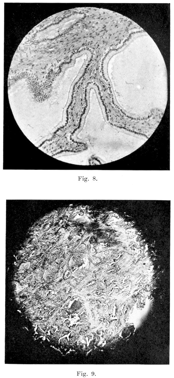
PLATE V
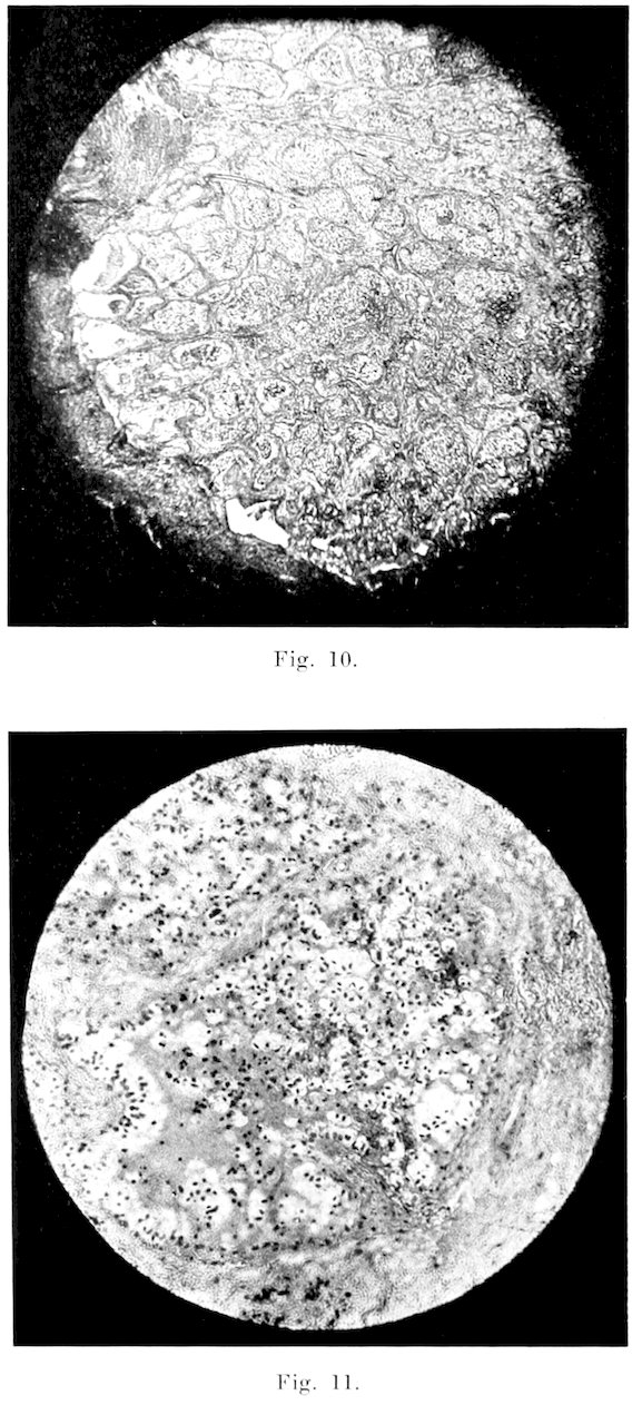
PLATE VI
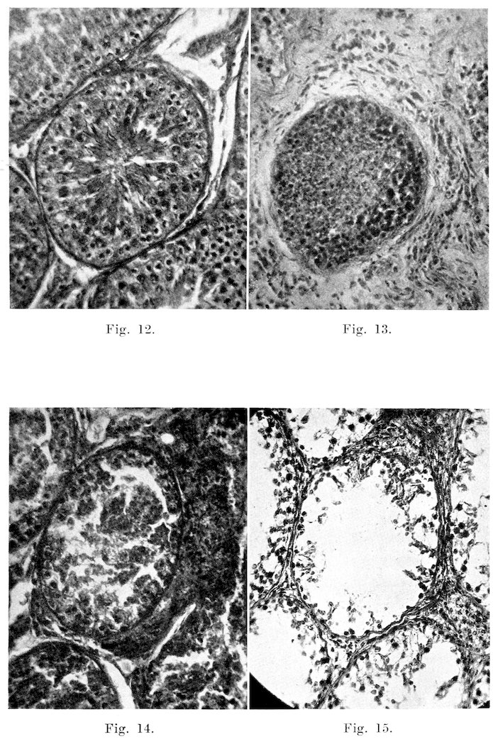
PLATE VII
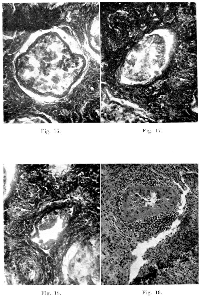
PLATE VIII
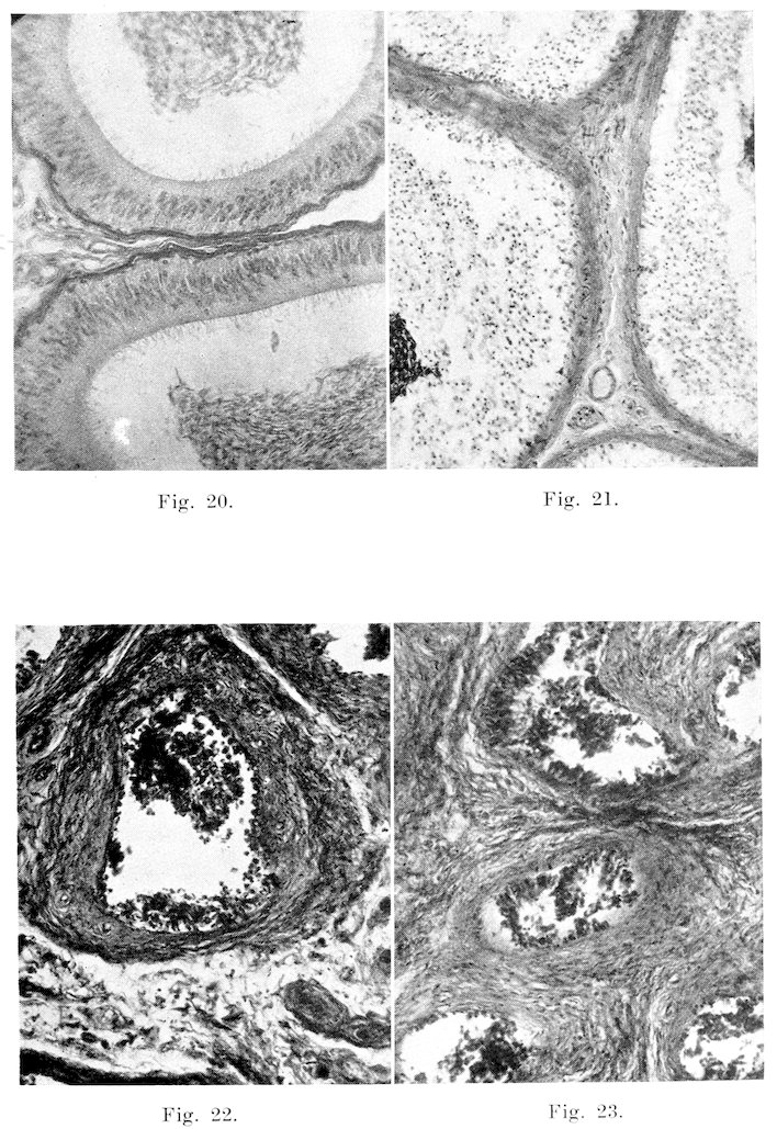
PLATE IX
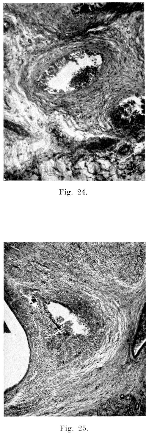
PLATE X
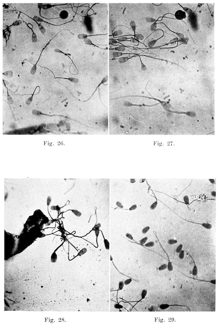
PLATE XI
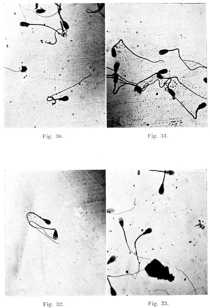
PLATE XII
TRANSCRIBER’S NOTES
Page Changed from Changed to
54 [blank] Bacteriology of the Genital Tracts of Mature Infertile or Sterile Bulls Group VI
Typos fixed; non-standard spelling and dialect retained.