← Table of contents
A New Species of Heteromyid Rodent from the Middle Oligocene of Northeast Colorado with Remarks on the Skull

EDWIN C. GALBREATH

Reading edition

University of Kansas Publications Museum of Natural History Volume 1, No. 18, pp. 285-300, 2 plates August 16, 1948 University of Kansas LAWRENCE 1948 University of Kansas Publications, Museum of Natural History Editors: E. Raymond Hall, Chairman; H. H. Lane, Edward H. Taylor Volume 1, No. 18, pp. 285-300, 2 plates August 16, 1948 University of Kansas Lawrence, Kansas PRINTED BY FERD VOILAND, JR., STATE PRINTER TOPEKA, KANSAS 1948 22-3342

Plate 2. Heliscomys tenuiceps. Univ. Kans. Mus. Nat. Hist., Vert. Paleo. Coll. No. 7702. A, dorsal view; B, lateral view; C, ventral view. All views approximately × 5.
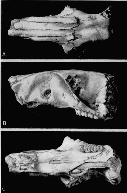
Plate 2. Heliscomys tenuiceps. Univ. Kans. Mus. Nat. Hist., Vert. Paleo. Coll. No. 7702. A, dorsal view; B, lateral view; C, ventral view. All views approximately × 5.

Plate 3. Heliscomys tenuiceps. Univ. Kans. Mus. Nat. Hist., Vert. Paleo. Coll. No. 7702. A, lateral view of right side of skull showing structures in orbital area. ALS, alisphenoid. FR, frontal. MAX, maxillary. OS, orbitosphenoid. PAL, palatine. PC, presphenoid canal. SF, sphenoidal fissure. SFr, sphenofrontal foramen. SPal, sphenopalatine foramen. Approximately × 9.3; B, occlusal view of P4-M3. Approximately × 23.4.
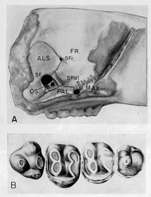
Plate 3. Heliscomys tenuiceps. Univ. Kans. Mus. Nat. Hist., Vert. Paleo. Coll. No. 7702. A, lateral view of right side of skull showing structures in orbital area. ALS, alisphenoid. FR, frontal. MAX, maxillary. OS, orbitosphenoid. PAL, palatine. PC, presphenoid canal. SF, sphenoidal fissure. SFr, sphenofrontal foramen. SPal, sphenopalatine foramen. Approximately × 9.3; B, occlusal view of P4-M3. Approximately × 23.4.
Explore with AITake your curiosity a little further.

Reading settings