Class—CRUSTACEA. Sub-Class—CIRRIPEDIA.
Reading edition
Family—LEPADIDÆ.
Cirripedia pedunculo flexili, musculis instructo: scutis[3] musculo adductore solummodô instructis: valvis cæteris, siquæ adsunt, in annulum immobilem haud conjunctis.
Cirripedia having a peduncle, flexible, and provided with muscles. Scuta[3] furnished only with an adductor muscle: other valves, when present, not united into an immovable ring.
Metamorphoses; larva, first stage, pp. 9-12; larva, second stage, p. 13; larva, last stage, p. 14; its carapace, ib.; acoustic organs, p. 15; antennæ, ib.; eyes, p. 16; mouth, p. 17; thorax and limbs, p. 18; abdomen, p. 19; viscera, ib.; immature cirripede, p. 20; homologies of parts, p. 25.
Description of mature Lepadidæ, p. 28; capitulum, ib.; peduncle, p. 31; attachment, p. 33; filamentary appendages, p. 38; shape of body, and muscular system, p. 39; mouth, ib.; cirri, p. 42; caudal appendages, p. 43; alimentary canal, 44; circulatory system, p. 46; nervous system, ib.; eyes, p. 49; olfactory organs, p. 52; acoustic(?) organs, p. 53; male sexual organs, p. 55; female organs, p. 56; ovigerous lamellæ, p. 58; ovigerous fræna, ib.; exuviation, p. 61; rate of growth, ib.; size, ib.; affinities of family, p. 64; range and habitats, p. 65; geological history, p. 66.
Metamorphoses.—I will here briefly describe the Metamorphoses, as far as known, common to all Cirripedia, but more especially in relation to the present family. I may premise, that since Vaughan Thompson’s capital discovery of the larvæ in the last stage of development in Balanus, much has been done on this subject: this same author subsequently published[4] in the ‘Philosophical Transactions,’ an account of the larvæ of Lepas and Conchoderma (Cineras) in the first stage; and seeing how totally distinct they were from the larva of the latter stage in Balanus, he erroneously attributed the difference to the difference in the two families, instead of to the stage of development. Burmeister[5] first showed, and the discovery is an important one, that in Lepas the larvæ pass through two totally different stages. This has subsequently been proved by implication to be the case in Balanus, by Goodsir,[6] who has given excellent drawings of the larva in the first stage; and quite lately, Mr. C. Spence Bate, of Swansea, has made other detailed observations and drawings of the larvæ of five species in this same early stage, and has most kindly permitted me to quote from his unpublished paper[7]. I am enabled to confirm and generalise these observations, in all the Cirripedes in the Order containing the Balanidæ and Lepadidæ.
The ova, and consequently the larvæ of the Lepadidæ, in the First Stage, whilst within the sack of the parent, vary in length from .007 to .009 in Lepas, to .023 of an inch in Scalpellum: my chief examination of these larvæ has been confined to those of Scalpellum vulgare; but I saw them in all the other genera. The larva is somewhat depressed, but nearly globular; the carapace anteriorly is truncated, with lateral horns; the sternal surface is flat and broad, and formed of thinner membrane than the dorsal. The horns just alluded to are long in Lepas and short in Scalpellum; their ends are either rounded and excessively transparent, or, as in Ibla, furnished with an abrupt, minute, sharp point: within these horns, I distinctly saw a long filiformed organ, bearing excessively fine hairs in lines, so exactly like the long plumose spines on the prehensile antennæ of the larvæ in the last stage; that I have not the least doubt, that these horns are the cases in which antennæ are in process of formation. Posteriorly to them, on the sternal surface, near each other, there are two other minute, doubly curved, pointed horns, about .004 in length, directed posteriorly; and within these I again saw a most delicate articulated filiformed organ on a thicker pedicel: in an excellent drawing, by Mr. C. S. Bate, of the larva of a Chthamalus (Balanus punctatus of British authors), after having kept alive and moulted once, these organs are distinctly shown as articulated antennæ (without a case), directed forwards: hence, before the first moult in Scalpellum, we have two pair of antennæ in process of formation. Anteriorly to the bases of these smaller antennæ is seated the heart-shaped eye, (as I believe it to be,) .001 in diameter, with apparently a single lens, surrounded, except at the apex, by dark-reddish pigment-cells. In some cases, as in some species of Lepas, the larvæ, when first excluded from the egg, have not an eye, or a very imperfect one.
There are three pairs of limbs, seated close together in a longitudinal line, but some way apart in a transverse direction: the first pair always consists of a single spinose ramus, it is not articulated in Scalpellum, but is multi-articulate in some genera; it is directed forwards. The other two pair have each two rami, supported on a common haunch or pedicel: in both pair, the longer ramus is multi-articulate, and the shorter ramus is without articulations, or with only traces of them: the longer spines borne on these limbs (at least, in Scalpellum and Chthamalus,) are finely plumose. The abdomen terminates, a little beyond the posterior end of the carapace, in a slightly upturned horny point; a short distance anteriorly to this point, a strong, spinose, forked projection depends from the abdominal surface.
Messrs. V. Thompson, Goodsir, and Bate, have kept alive for several days the larvæ of Lepas, Conchoderma, Balanus, Verruca, and Chthamalus, and have described the changes which supervene between the first and third exuviations. The most conspicuous new character is the great elongation of the posterior point of the carapace into an almost filiform, spinose point in Lepas, Conchoderma, Chthamalus, and Balanus, but not according to Goodsir, in one of the species of the latter genus. The posterior point, also, of the abdomen becomes developed in Balanus (Goodsir) into two very long, spear-like processes, serrated on their outer sides; in Lepas and Conchoderma, according to Thompson, into a single, tapering spinose projection; and in Chthamalus, as figured by Mr. Bate, the posterior bifid point, as well as the depending ventral fork, increase much in size. Another important change, which has been particularly attended to by Mr. Bate, is the appearance of spinose projections and spines (some of which are thick, curved, and strongly plumose, or, almost pectinated along their inner sides) on the pedicels and lower segments of the shorter rami of the two posterior pairs of limbs.
The mouth in its earliest condition alone remains to be described; in S. vulgare, it is seated on a very slight prominence, in a most remarkable situation, namely, in a central point between the bases of the three pairs of legs. I traced by dissection the œsophagus for some little way, until lost in the cellular and oily matter filling the whole animal, and it was directed anteriorly, which is the direction that might have been expected, from the course followed by the œsophagus in the larva in the last stage, and in mature Cirripedes. Mr. A. Hancock has called my attention to a probosciformed projection on the under side of the larva of Lepas fascicularis, when just escaped from the egg. Mr. Bate has described this same proboscis in Balanus and Chthamalus, and states the important fact, that it is capable of being moved by the animal; and, lastly, I have seen it in an Australian Chthamalus, and in Ibla, of remarkable size. This proboscis, which is always directed posteriorly, (like the mouth in the mature animal,) certainly answers to the mouth as made out by dissection in Scalpellum; and I believe I saw, as has Mr. Bate, a terminal orifice: it certainly does not possess any trophi. In Ibla (in which the larva is large enough for dissection), the base of the proboscis arises posteriorly to the first pair of legs, and the orifice at the other end reaches beyond or posteriorly to the point, where the mouth in Scalpellum opens, namely between the middle pair of legs. The mouth being either so largely probosciformed or seated only on a slight eminence, in two genera so closely allied as Ibla and Scalpellum, and (judging from Mr. Thompson’s figures, and from what I have seen myself,) in the species of the same genus Lepas, is a singular difference: in the cases in which, at first, the proboscis is absent, it would probably soon be developed. I cannot but suppose that the inwardly directed spines on the bases of the two posterior legs, which are so rapidly developed, serve some important end, namely, as organs of prehension for the larvæ, like the mandibles and maxillæ of mature Cirripedes, for seizing their prey, and conveying it to their moveable mouths, conveniently seated for this purpose.
The first pair of legs answers, as I believe from reasons hereafter to be assigned, to the outer pair of maxillipods in the higher crustacea; and the other four legs to the first two pair of thoracic limbs in these same crustacea; this being the case, the highly remarkable position of the mouth in the larva, either between the bases of the two posterior pair of legs, or at least posteriorly to the first pair, together with the probable functions of the spiny points springing from the basal segments of the two hinder pair of true thoracic limbs, forcibly bring to mind the anomalous structure of the mouth being situated in the middle of the under side of the thorax, in Limulus,—that most ancient of crustaceans, and therefore one likely to exhibit a structure now embryonic in other orders. I will only further remark, that I suspect that the truncation of the anterior end of the carapace, has been effected by the segments having been driven inwards, and consequently, that the larger antennæ within the lateral horns, though standing more in front than the little approximate pair, are normally the posterior of the two pair. According to Milne Edwards, the posterior pair are normally seated outside the anterior pair, and this is the case with those within the lateral horns.
Larva in the Second Stage.—Notwithstanding the considerable changes, already briefly given, which the larva undergoes during the first two or three exuviations after leaving the egg, all these forms may be conveniently classed under the first stage. The larva in the Second stage is known only from a single specimen described, figured, and found by Burmeister,[8] adhering to sea-weed in the midst of other larvæ of Lepas in the last stage. In its general shape and compressed form, it seems to come nearer to the last than to the first stage. It has only three pair of legs, situated much more posteriorly on the body than in the first stage, and all directed posteriorly; they are much shorter than heretofore, and resemble rather closely those of the last stage, with the important exception that the first pair has only one ramus. It is this circumstance which leaves no doubt on my mind, that we here have the three pair of limbs, of the first stage, metamorphosed. The body is prolonged some way behind these limbs, and ends in a blunt, rounded point, in which, probably, are developed the three posterior pair of legs and the abdomen of the larva in the last stage. The mouth is now seated some way anteriorly to the limbs, is large and probosciformed, and is, I presume, still destitute of trophi. There are now two closely approximate eyes, but as yet both are simple. The smaller pair of antennæ has disappeared. The whole animal was attached to the sea-weed by a (I presume, pair of,) “fleischigen Fortsatz,” which Burmeister considers as the prehensile antennæ, to be presently described, in an early state of development. I have little doubt that this is correct, for in an abnormal Cirripede of another order, in which the larva appears in the first stage with prehensile antennæ, the eggs have two great projecting horns including these organs, and attached by their tips, through some unknown means, to the sack of the parent, apparently in the same manner as Burmeister’s larva was attached to the sea-weed. I will only further remark on the larva of this Second stage, that its chief development since the first stage, has been towards its anterior end. The next great development, to be immediately described, is towards the posterior end of the animal.
Larva, Last Stage.—My chief examination has been directed, at this stage of development, to the larvæ of Lepas australis, which are of unusual size, namely, from .065 to even almost .1 of an inch in length; I examined, however, the larvæ of several other species of Lepas, of Ibla and of Balanus, with less care, but sufficiently to show that in all essential points of organisation they were identical; this, indeed, might have been inferred from the similarity of the larval prehensile antennæ, preserved in the bases of all mature Cirripedes, and which I have carefully inspected in almost every genus. The larvæ in this final stage, in most of the genera, have increased many times in size since their exclusion from the egg; for instance, in Lepas australis, from .007 to .065, or even to .1 of an inch. They are now much compressed, nearly of the shape of a cypris or mussel-shell, with the anterior end the thickest, the sternal surface nearly or quite straight, and the dorsal arched. Almost the whole of what is externally visible consists of the carapace; for the thorax and limbs are hidden and enclosed by its backward prolongation; and even at the anterior end of the animal, the narrow sternal surface can be drawn up, so as to be likewise enclosed. As in several Stomapod crustaceans, the part of the head bearing the antennæ and organs of sense, in front of the mouth, equals, or even exceeds in length, and more than exceeds in bulk, the posterior part of the body, consisting of the enclosed thorax and abdomen. I will now briefly describe, in the following order, the carapace, the organs of sense, mouth, thorax and limbs, abdomen, and internal viscera.
The form of the Carapace has been sufficiently described; it consists of thick chitine membrane, marked with lines, and sometimes with stars and other patterns; it is obscurely divided into two halves by a line or suture along part of the dorsal margin; these halves or two valves are drawn together by an adductor muscle, in the same relative position as in the mature Cirripede. The part overhanging and enclosing the thorax is lined by an excessively delicate membrane, obviously homologous with the lining of the sack in the mature animal, and is nothing but a duplicature of the carapace, rendered very thin from being on the under or protected side: a layer of true skin or corium, probably double, separates these two folds.
Acoustic Organs.—On the borders of the carapace, at the anterior end, on the sternal surface, there are two minute orifices, in L. australis .002 in diameter, sometimes having a distinct border round them; the membrane of the carapace on the inside is prolonged upwards and inwards in two short funnel-shaped tubes, lodged in closed sacks of the corium: within these sacks on each side a delicate bag is suspended, and hangs in the mouth of the above funnel; at the upper end a large nerve could be distinctly seen to enter the bag: I cannot doubt that this is a sense-organ; from its position and from the animal not feeding (as we shall presently see), I conclude that it is an acoustic organ.
Antennæ.—These are large and conspicuous; they are attached very obliquely on the sternal surface, a little way from the anterior end of the carapace, beyond which, when exserted, they extend;[9] they can (at least in Ibla) be retracted within the carapace. They consist of three segments: the first or basal one is much larger than the others, and apparently always has a single spine on the outer distal margin. The second segment consists either of a large, thin, circular, sucking disc, or is hoof-like (Tab. V, figs. 5, 10, 11, 12); in all cases it is furnished with one or more spines, (seven very long ones in Lepas,) on the exterior-hinder margin. The third and ultimate segment is small; it is articulated on the upper surface of the disc, and is directed rectangularly outwards; it is sometimes notched, and even shows traces of being bifid; it bears about seven spines at the end; some of these spines are hooked, others simple, and in Lepas and Conchoderma, two or three are very long, highly flexible, and plumose, a double row of excessively fine hairs being articulated on them. I can hardly doubt that these latter spines, (within which the purple corium could be seen to enter a little way,) floating laterally outwards, serve as feelers. The antennæ, at first, are well furnished with muscles. They serve, in Lepas, according to Mr. King, and in Balanus, according to Mr. Bate, and as I saw myself in another unnamed order, for the purpose of walking, one limb being stretched out before the other; but their main function is to attach the larva for its final metamorphosis into a Cirripede. The disc can adhere even to so smooth a surface as a glass tumbler.[10] The attachment is at first manifestly voluntary, but soon becomes involuntary and permanent, being effected by special and most remarkable means, which will be most conveniently described in a later part of this Introduction. I will here only state that I traced with ease the two cement-ducts running from two large glandular bodies, to within the antennæ up to the discs.
Eyes.—Close behind the basal articulations of the antennæ, the sternal surface consists of two approximate, elongated, narrow, flat pieces, or segments. These Burmeister considers as the basal segments of the antennæ: as they are not cylindrical, I do not see the grounds for this conclusion: their posterior ends are rounded, and the membrane forming them is reflected inwards, in the form of two, forked, horny apodemes, together resembling two letters, UU, close together; these project up, inside the animal, for at least one third of its thickness from the sternal to the dorsal surface. The two great, almost spherical eyes in L. australis, each 1/150th of an inch in diameter, are attached to the outer arms, thus, °UU°, in the position of the two full stops. Hence the eyes are included within the carapace. Each eye consists of eight or ten lenses, varying in diameter in the same individual from 1/2000 to 3/2000th of an inch, enclosed in a common membranous bag or cornea, and thus attached to the outer apodemes. The lenses are surrounded half way up by a layer of dark pigment-cells. The nerve does not enter the bluntly-pointed basal end of the common eye, but on one side of the apodeme. The structure here described is exactly that found, according to Milne Edwards, in certain crustacea. In specimens just attached, in which no absorption has taken place, two long muscles with transverse striæ may be found attached to the knobbed tips of the two middle arms of the two °UU°, and running up to the antero-dorsal surface of the carapace, where they are attached; other muscles (without transverse striæ) are attached round the bases, on both sides of both forks. The action of these muscles would inevitably move the eyes, but I suspect that their function may be to draw up the narrow, deeply folded, sternal surface, and thus cause the retraction of the great prehensile antennæ within the carapace.
Mouth.—This is seated in exactly the same position as in the mature Cirripede, on a slight prominence, fronting the thoracic limbs, and so far within the carapace, that it was obviously quite unfitted for the seizure of prey; and it was equally obvious, that the limbs were natatory, and incapable of carrying food to the mouth. This enigma was at once explained by an examination of the mouth, which was found to be in a rudimentary condition and absolutely closed, so that there would be no use in prey being seized. Underneath this slightly prominent and closed mouth, I found all the masticatory organs of a Cirripede, in an immature condition. The state of the mouth will be at once understood, if we suppose very fluid matter to be poured over the protuberant mouth of a Cirripede, so as to run a little way down, in the shape of internal crests, between the different parts, and in the shape of a short, shrivelled, certainly closed tube, a little way (.008 of an inch in L. australis) down the œsophagus. Hence, the larva in this, its last stage, cannot eat; it may be called a locomotive Pupa;[11] its whole organisation is apparently adapted for the one great end of finding a proper site for its attachment and final metamorphosis.
Thorax and Limbs.—The thorax is much compressed, and consists of six segments, corresponding with the six pair of natatory legs; the anterior segments are much plainer (even the first being distinctly separated by a fold from the mouth), than the posterior segments, which is exactly the reverse of what takes place in the mature Cirripede; in the latter, the first segment is confounded with the part bearing the mouth. The epimeral elements of the thorax are distinguishable; the sternal surface is very narrow, and is covered with complicated folds and ridges. The six pair of legs are all close, one behind the other, and all are alike in having a haunch or pedicel of two segments, directed forwards, bearing two arms or rami, each composed of two segments, the outer ramus being a little longer than the inner one. On the lower segments in both rami of all the limbs, there is a single spine. In all the limbs, the obliquely truncated summit of the terminal segment of the inner ramus bears three very long, beautifully plumose spines: in the first pair, the summit of the outer ramus bears four, and in the five succeeding pair, six similar spines. This difference, small as it is, is interesting, as recalling the much greater difference between the first and succeeding pairs, in the first and second stage of development. The terminal segments of all the rami, bearing the long plumose spines, are directed backwards. The limbs and thorax are well furnished with striated muscles. The animal, according to Mr. King, swims with great rapidity, back downwards. The limbs can be withdrawn within the carapace.
Abdomen and Caudal Appendages.—The abdomen is small, and its structure might easily be overlooked without careful dissection of the different parts: it consists of three segments; the first can be seen to be distinct from the last thoracic segment, bearing the sixth pair of limbs, only from the fold of the epimeral element, and from its difference in shape; the second segment is very short, but quite distinct; the third is four or five times as long as the second, and bears at the end two little appendages, each consisting of two segments, the lower one with a single spine, and the upper one with three, very long, plumose spines, like those on the rami of the thoracic limbs. The abdomen contains only the rectum and two delicate muscles running into the two appendages, between the bases of which the anus is seated.
Internal Viscera.—Within the body, in front of the mouth, it was easy to find the stomach (with two pear-shaped cæca at the upper end), running first anteriorly, and then curving back and reaching the anus by a long rectum, difficult to be followed: it appeared, however, to me, that this stomach had more relation to the young Cirripede, of which every part could now generally be traced, than to the larva, with its closed and rudimentary mouth: the fact, however, of its being prolonged to the anus, which is in a different position in the larva and mature state, shows that the stomach serves, at least, as an excretory channel. Besides the stomach, the several muscles already alluded to, and much pulpy and oily matter, the only other internal organs consist of two long, rather thick, gut-formed masses, into the anterior ends of which the cement-ducts running from the prehensile antennæ could be traced. These masses are formed of irregular orange balls, about .001 of an inch in diameter, made up of rather large cells, so to have a grape-like appearance, held together by a transparent pale yellowish substance, but apparently not enclosed in a membrane: these masses lie rather obliquely, and approach each other at their anterior ends; they extend from above the compound eyes, to the cæca of the stomach to which they cohere, but in young specimens, they extend some way beyond the cæca, between the folds of the carapace. The two cement-ducts, at the points where they enter these bodies, expand and are lost; at this point, also, the little orange-coloured masses of cells have the appearance of being broken down into a finer substance. Within the cement-ducts I saw a distinct chord of rather opaque cellular matter. We shall presently see, that these gut-formed masses are the incipient ovaria.
The Young Cirripede within the Larva.—Several times I succeeded in dissecting off the integuments of the lately-attached larva, and in displaying the young Lepas australis entire. The following description applies to the Cirripede in this state; but for convenience sake, I shall occasionally refer to its condition when a little more advanced. I may premise, and the fact in itself is curious, that the bivalve-like shell of the larva, together with the compound eyes, is first moulted, and some time afterwards, the inner lining of the sack, together with the integuments of the thorax and of the natatory legs: hence, I often found specimens, which externally seemed to have perfected their metamorphoses, but which, within their sacks, retained all the characters of the natatory larva. According to Mr. King, the larva of Lepas throws off its external shell five days after becoming attached. Whilst the young Lepas is closely packed within the larva, the capitulum, as known by the five valves, about equals in length the peduncle. The peduncle occupies the anterior half of the larva; when fully stretched, it becomes narrower and slightly longer than the capitulum; the separation between the capitulum and peduncle is almost arbitrary in the mature animal, and corresponds with no particular line in the larva. Even at this early period, the muscles of the peduncle are quite distinct. No vestige is preserved in the outer integument, of the sternal and dorsal sutures of the larval carapace; but in the corium of the peduncle, three coloured marks which occur near the eyes, and two little curled marks which occur near the acoustic orifices of the larva, are all preserved for some time after maturity. The compound eyes, as we have seen, are attached to apodemes, springing from the sternal surface of the larval carapace, and are consequently cast off with it: whilst the young Cirripede is packed within the larva, the outer integument of its peduncle necessarily forms a deep transverse fold passing over the eyes and apodemes, and this, as we shall presently see, plays an important part in the future position of the animal. The antennæ are not moulted with the carapace, but left cemented to the surface of attachment; their muscles are converted into sinewy fibres, the corium after a short period is absorbed, and they are then preserved in a functionless condition. No trace of the two acoustic sacks can be perceived in the corium of the young Cirripede, excepting the coloured marks above alluded to.
In the young capitulum, the five valves stand some way apart from each other; they are elegant objects under the microscope; they are not calcified, but consist exclusively of chitine; they are rather thick, composed of an outer membrane lined by hexagonal prisms, quite unlike any other membrane in the animal. These valves, which I have called primordial valves, resemble pretty closely in shape the valves of the mature animal; the fork of the carina, however, is indicated only by a slight constriction above the lower end. After the exuviation of the larval integuments, and when calcification commences, the first layer of shell is deposited under, and then round these primordial valves. The latter, in well preserved old specimens, may often be detected on the umbones of the scuta, terga, and carina, but not on the umbones of any other valves.
The mouth seems one of the earliest parts developed: in the youngest larva dissected, I could make out at least points corresponding with each organ; and, at the period when the young Cirripede could be dissected out of its larval envelopes, their general details were quite plain. The labrum, however, had not become bullate. The mouth, as we have seen, is formed under the rudimentary mouth of the larva, and at the same relative spot occupied by the probosciformed mouth of the larva in the second stage. Thus far, in the young Cirripede and larva, there has been no great change in the relative positions of the parts: the rudimentary eyes, however, of the former are developed posteriorly to (or above, as applied to a Cirripede,) the cast-off compound eyes of the larva; but the position of the mouth, of the antennæ, and of the several coloured marks in the corium, prove to demonstration, the correspondence in both of part to part. The case is rather different with what follows.
The Cirri are developed at first of considerable length, so that the young animal may soon provide itself with food; in Lepas australis they are of great length, the sixth pair consisting of seventeen or eighteen obscure segments. The extreme tips of the twenty-four rami of the six pair of cirri, are formed within the twenty-four, corresponding, little, bi-segmental rami of the six pair of natatory legs; but as the cirri are many times longer than these legs, they occupy in a bundle the whole thorax of the larva; no part whatever of the thorax of the Cirripede is formed within the thorax of the larva, but (together with the pedicels of the anterior cirri) within the cephalic cavity. As a consequence of this, the longitudinal axis of the thorax of the young Cirripede lies almost transversely to the longitudinal axis of the larva; and the Cirripede, from this transverse position of its thorax, comes to be, as it were, internally, almost cut in twain, and the sack thus produced. As soon as the young Cirripede is free and can move itself, the cirri are curled up, and the thorax is advanced towards the orifice of the capitulum, its longitudinal axis resuming the position of approximate parallelism to the longitudinal axis of the whole body, which it had in the larval condition. The reader will, perhaps, understand what I mean, if he will look at the mature Cirripede, figured in Pl. IX, fig. 4. In this, he will see that the body or thorax is united to the peduncle only by a small part below the mouth; on the other hand, if he imagines the whole bottom of the body (as high up as the letter h) united and blended into the peduncle, he will see the state in which these parts exist in the larva. Now, let him greatly shorten the cirri, so as to resemble the natatory legs of the larva, and then imagine a young Cirripede, with cirri of full length, formed within the old one, he will see that the new thorax supporting the cirri will have to be developed in an almost transverse position,—the animal consequently being internally almost separated into twain.
Of the internal organs, whilst the Cirripede is still within the larva, I have already mentioned the stomach with its pair of cæca: from the retracted position of the thorax and rudimentary abdomen, and consequently of the anus, compared with these parts in the larva, the alimentary canal is not above half its former length. There is, as yet, no trace of the filaments supposed by some to act as branchiæ, at the base of the first pair of cirri. Nor could I perceive a trace of the testes or vesiculæ seminales: the penis is represented by a minute, apparently imperforate projection. I have already briefly described the pair of large, gut-formed bodies in the larva, into the anterior ends of which the cement-ducts ran, and evidently derived their slightly opaque, cellular contents. At a very early age, before the young Cirripede can be distinctly made out, the posterior ends of these gut-formed bodies are absorbed, so as not to pass beyond the cæca of the stomach. When the young Cirripede is plainly developed within the larva, these bodies in a relatively reduced condition are still distinct near the cæca, and at the opposite or anterior end (i. e. lower, in the position in which Cirripedes are usually figured), they have branched out into a sheet of delicate inosculating tubes; these could be traced by every stage, until, in the young perfected Cirripede, they filled the peduncle as ordinary ovarian tubes. In the larva, the two gut-formed bodies or incipient ovaria keep of equal thickness from one to the other end, but in the mature Cirripede, the ovarian tubes in the peduncle and the small, glandular, grape-like masses, near the stomach-cæca, are connected only by a delicate tube; this I failed in tracing in specimens in the very immature condition of those now under description.
The larva fixes itself with its sternal surface parallel and close to the surface of attachment, and the antennæ become cemented to it: if the Cirripede, after its metamorphosis had remained in this position, the cirri could not have been exserted, or only against the surface of attachment; but there is a special provision, that the young Cirripede shall immediately assume its proper position at right angles to the position which it held whilst within the larva, namely with its posterior end upwards. This is effected in a singular manner by the exuviation of the great compound eyes, which we have seen are fastened to the outer arms of the double °UU°-like, sternal apodemes: these together with the eyes stretch transversely across, and internally far up into, the body of the larva; and, as the whole has to be rejected or moulted, the membrane of the peduncle of the young Cirripede has necessarily to be formed with a wide and deep inward fold, extending transversely across it; this when stretched open, after the exuviation of the larval carapace and apodemes, necessarily causes the sternal side of the peduncle to be longer than the dorsal, and, as a consequence, gives to the young Cirripede its normal position, at right angles to that of the larva when first attached.
I may here state, that I have examined the larvæ in this the final or perfect stage in four species of Lepas, in Conchodermavirgata, Ibla quadrivalvis, and, though rather less minutely, in Balanus balanoides, and I find all essential points of organisation similar. With the exception of diversities in the proportional sizes of the different parts, and in the patterns on the carapace, the differences, even in the arrangement of the spines on the limbs and antennæ, are less than I should have anticipated.
I have in this abstract treated the metamorphoses at greater length than I should otherwise have done, on account of the great importance of arriving at a correct homological interpretation of the different parts of the mature animal. In Crustacea, according to the ordinary view, there are twenty-one segments; of these I can recognise in the Cirripede, on evidence as good as can generally be obtained, all with the exception of the four terminal abdominal segments; these do not occur in any species known to me, in any stage of its development. If that part of the larva in front of the mouth, bearing the eyes, the prehensile antennæ, and in an earlier stage two pair of antennæ, be formed, as is admitted in all other Crustacea, of three segments, then beyond a doubt, from the absolute correspondence of every part, and even every coloured mark, the peduncle of the Lepadidæ is likewise thus formed. The peduncle being filled by the branching ovarian tubes is no objection to this view, for I am informed on the high authority of Mr. J. D. Dana,[12] that this is the case with the cephalo-thorax in some true Crustaceans, for instance, in Sapphirina. To proceed, the mouth, formed of mandibles, maxillæ, and outer maxillæ, correspond with the fourth, fifth, and sixth segments of the archetype Crustacean. Posteriorly to the mouth, we come, in the larva, to a rather wide interspace without any apparent articulation or organ, and then to the thorax, formed of six segments, bearing the six pair of limbs, of which the first pair differs slightly from the others. The thorax is succeeded by three small segments, differently shaped, with the posterior one alone bearing appendages; these segments, I cannot doubt, from their appearance alone, and from their apparent function of steering the body, are abdominal segments. If this latter view be correct, the thoracic segments are the six posterior ones of the normal seven segments, and there must be two segments missing between the outer maxillæ and first thoracic pair of legs, which latter on this view springs from the ninth segment. Now, in a very singular Cirripede, already alluded to under the name of Proteolepas, the two missing segments are present, the mouth being actually succeeded by eight segments, and these by the three usual abdominal segments,—every segment in the body being as distinct as in an Annelid: hence in Proteolepas, adding the three segments for the mouth and three for the carapace, we have altogether seventeen segments, which, as I stated, is the full number ever observed in any Cirripede, the four missing ones being abdominal, and, I presume, the four terminal segments. That the cavity in which the thorax is lodged, in the larva and therefore in the mature Cirripede, is simply formed by the backward production of the carapace, does not require any discussion. The valves have no homological signification.
As we have just seen that the first pair of natatory legs is borne on the ninth segment of the body, so it must be with the first pair of cirri, which consequently correspond to the outer maxillipods (the two inner pair of maxillipods or pied-machoires being here aborted) of the higher Crustacea, and hence their difference from the five posterior pair, which correspond with the five, ordinary pair of ambulatory legs in these same Crustacea. The part of the body, which I have called the prosoma, that is the protuberant, non-articulated, lower part of the thorax (Pl. IX, fig. 4 n), is a special development, either of the ninth segment, bearing the first pair of cirri, or of the segments corresponding with the organs of the mouth. The three abdominal segments of the larva are represented in the mature Cirripede, in the Order containing the Lepadidæ, only by a minute, triangular gusset, let in between the V-shaped tergal arches of the last thoracic segment: in this gusset, small as it is, is seated the anus, and on each side the caudal appendages, often rudimentary and sometimes absent. In another order, I may remark, (including, probably, the Alcippe of Mr. Hancock,) the cirri, of which there are only three pair, are abdominal.
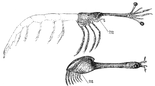
I feel much confidence, that the homologies here given are correct. The cause of their having been generally overlooked arises, I believe, from the peculiar manner, already described, in which the animal, during its last metamorphosis, is internally almost intersected: even for some little time after discovering that the larval antennæ were always embedded in the centre of the surface of attachment, I did not perceive, that this was the anterior end of the whole animal. The accompanying woodcut gives at a glance, a view of the homologies of the external parts: the upper figure (from Milne Edwards) is a Stomapod Crustacean, Leucifer of Vaughan Thompson, and the abdomen, which we know becomes in Cirripedes, after the metamorphosis, rudimentary, and therefore does not fairly enter into the comparison, is given only in faint lines: the lower figure is a mature Lepas, with the antennæ and eyes, which are actually present in the larva, retained and supposed to have gone on growing. All that we externally see of a Cirripede, whether pedunculated or sessile, is the three anterior segments of the head of a Crustacean, with its anterior end permanently cemented to a surface of attachment, and with its posterior end projecting vertically from it.
[m.—Mouth.]
CAPITULUM.
I will now proceed to a general description of the different parts and organs in the Lepadidæ. The Capitulum is usually much flattened, but sometimes broadly oval in section. It is generally formed of five or more valves, connected together by very narrow or broad strips of membrane; sometimes the valves are rudimental or absent, when the whole consists of membrane. When the valves are numerous, and they occasionally exceed a hundred in number, they are arranged in whorls, with each valve generally so placed as to cover the interval between the two valves above. Of all the valves, the scuta are the most persistent; then come the terga, and then the carina; the rostrum and latera occur only in Scalpellum and Pollicipes, and in a rudimentary condition in Lithotrya, and, perhaps, in the fossil genus Loricula. The valves are formed sometimes of chitine (as in Ibla and Alepas), but usually of shell, which varies from transparency to entire opacity. The shell is generally white, occasionally reddish or purple; exteriorly, the valves are covered by more or less persistent, generally yellow, strong membrane. The scuta and terga are always considerably larger than the other valves: in the different genera the valves differ so much in shape that little can be predicated of them in common; even the direction of their lines of growth differs,—thus, in Lepas and some allied genera, the chief growth of the scuta and of the carina is upwards, whereas in Pollicipes and Lithotrya, it is entirely downwards; in Oxynaspis, and some species of Scalpellum, it is both upwards and downwards. Even in the same species, there is often very considerable variation in the exact shape of the valves, more especially of the terga. The adductor muscle is always attached to a point not far from the middle of the scuta, and it generally has a pit for its attachment. In several genera, namely, Pæcilasma, Dichelaspis, Conchoderma, and Alepas, the scuta show a tendency to be bilobed or trilobed. The valves are placed either at some distance from each other, or close together; but their growing margins very rarely overlap each other, though this is sometimes the case with their upper, free, tile-like apices; in a few species the scuta and terga are articulated together, or united by a fold. The membrane connecting the valves, where they do not touch each other, is like that forming the peduncle, and is sometimes brilliantly coloured crimson-red; generally, it appears blueish-gray, from the corium being seen through. Small pointed spines, connected with the underlying corium by tubuli, are not unfrequently articulated on this membrane: the tubuli, however, are often present where there are no spines. To allow of the growth of the capitulum, the membrane between the valves splits at each period of exuviation, when a new strip of membrane is formed beneath, connected on each side with a fresh layer of shell,—the old and outer slips of membrane disintegrating and disappearing: when there are many valves, the line of splitting is singularly complicated. This membrane consists of chitine,[13] and is composed of numerous fine laminæ. After the valves have been placed in acid, a residue, very different in bulk in different genera, is left, also composed of successive laminæ of chitine. It appears to me that each single lamina of calcified chitine, composing the shell, must once have been continuous with a non-calcified lamina in the membrane connecting the several valves: at the line where this change in calcification supervenes, the chitine generally assumes some colour, and becomes much harder and more persistent; and as the whole valve is formed of component laminæ thus edged (the once continuous laminæ of non-calcified chitine connecting the valves, having disintegrated and disappeared) the surfaces of the valves are generally left covered by a persistent membrane, constituted of these edgings: this membrane has been called the epidermis. In some genera, as in Lepas, this so-called epidermis is seldom preserved, excepting on the last zone of growth: in Scalpellum and Pollicipes it usually covers the whole valves. It appears to me that the laminæ of chitine, and of calcified chitine composing the valves, are both formed not by secretion, but by the metamorphosis of an outer layer of corium into these substances.
Within the capitulum is the sack, which, together with the upper internal part of the peduncle, encloses the animal’s body. The sack is lined by a most delicate membrane of chitine, under which there is a double layer of corium; this double layer is united together by short, strong, transverse bundles of fibres, branched at both ends:[14] in some genera, the ovarian tubes extend between these two layers. We have seen, under the head of the Metamorphoses, that the delicate tunic lining the sack is simply a duplicature of the thick membrane and valves forming the capitulum, the whole being the posterior portion of the carapace of the larva slightly modified.
Peduncle.—Its length varies greatly in different species, and even in the same species, according to the situation occupied by the individual; its lower end is sometimes pointed, but generally only a little narrower than the upper end. In outline, the peduncle is usually flattened, but sometimes quite cylindrical. It is composed of very strong, generally thick, transparent membrane, rarely coloured reddish, and often penetrated by numerous tubuli. The underlying corium is sometimes coloured in longitudinal bands. At each period of growth a new and larger integument is formed under the old one, which gradually disintegrates and disappears; the extreme lower point is often deserted by the corium, and ceases to grow, whilst the whole upper part still continues increasing in diameter: in length the chief addition is made (as is clearly seen in those genera having calcified scales), round the upper margin, at the base of the capitulum. The surface of the membrane is either naked or superficially clothed with minute, pointed, articulated spines, or it is penetrated by calcified scales or styles, (in Ibla alone formed of chitine,) which pass through it to the corium, and are added to at their bases, like the valves, at each period of growth. In Lithotrya alone the scales of the peduncle are moulted together with the connecting membrane. These scales on the peduncle are generally placed symmetrically in whorls, with each scale corresponding with the junctions of two scales, both above and below. Except in Scalpellum ornatum and the fossil Loricula pulchella, they are very small compared with the valves of the capitulum. When the scales are symmetrical, new ones are first formed only round the summit of the peduncle, and only those in the few uppermost whorls continue to grow or to be added to at their bases; afterwards membrane is deposited under them. The shelly matter of the scales resembles that of the valves, and the manner of growth is the same; tubuli generally run to and through them from the corium. From the continued enlargement of the membrane of the peduncle, the scales come to stand, in the lower portion, some way apart. In Ibla, new horny styles are formed indifferently in all parts of the peduncle. In some species of Pollicipes, the calcareous styles are not symmetrical or symmetrically arranged; and besides those first formed round the top of the peduncle, there are other and larger ones formed near its base. Lastly, in Lithotrya we have a row of calcareous discs or an irregular, basal cup, formed in the same manner as the valves of the capitulum: in this genus alone (as already stated,) the calcified scales are moulted, and here alone their edges are serrated.
The peduncle is lined within by three layers of muscles, longitudinal, transverse, and oblique, all destitute of the transverse striæ, characteristic of voluntary muscles; they run from the bottom of the peduncle to the base of the capitulum, as in Lepas, or half way up it, as in Conchoderma; in Alepas alone they surround the whole capitulum up to its summit. In Lithotrya there are two little, fan-like, transverse muscles (involuntary), extending from the basal points of the terga to a central line on the under side of the carina. The gentle swaying to and fro movements, and the great power of longitudinal contraction,—movements apparently common, as I infer from facts communicated to me by Mr. Peach, to all the Pedunculata,—are produced by these muscles. The interior of the peduncle is filled up with a great mass of branching ovarian tubes; but in Ibla and Lithotrya, the upper part of the peduncle is occupied by the animal’s body.
Means of Attachment.—If the peduncle be very carefully removed (Tab. IX, fig. 7 and Tab. I, fig. 6 b), from the surface of attachment, quite close to the end, but not at the actual apex, the larval prehensile antennæ can always be found: these have been sufficiently described for our present purpose under the head of the Metamorphoses; but I may add, that the diagnostic differences between them in the several genera are briefly given, for a special purpose, in a discussion on the sexes of Scalpellum at the end of that genus. We have seen in the larva, that the cement-ducts, with their opaque cellular contents, can be traced from within the discs of the antennæ to the anterior or lower ends of the two gut-formed bodies, which it can be demonstrated are the incipient ovaria.
In mature Cirripedes these ducts can be followed, in a slightly sinuous course, along the muscles on each side within the peduncle, till they expand into two small organs, which I have called cement-glands. These glands are found with great difficulty, except in Conchoderma aurita, where they are placed on each side under the inner layer of corium, at the bottom of the sack, so as to be just above the top of the peduncle; they resemble in shape a retort, (Pl. IX, fig. 3.). In Pollicipes mitella and polymerus they lie half way down the peduncle, close together, and apparently enclosed within a common membrane; in these two species the broad end of the gland is bent towards the neck of the retort. In Scalpellum the position is the same, but the shape is more globular. In Ibla the structure is more simple, namely, a tube slightly enlarged, running downwards, bent a little upwards, and then resuming its former downward course, the lower portion forming the duct. The gland contains a strongly coherent, pulpy, opaque, cellular mass, like that in the cement-ducts; but in some instances, presently to be mentioned, this cellular mass becomes converted within either the ducts or gland, or within both, into transparent, yellow, tough cement. Generally in Conchoderma, Pollicipes, and Scalpellum, two ovarian tubes, but in one specimen of Conchoderma aurita, three tubes, and in Ibla one tube could be seen running into or forming the gland; of the nature of the tubes there could not be the least doubt, for at a little distance from the glands they gave out branches (Pl. IX, fig. 3), containing ova in every state of development. In some specimens as in that figured of Conchoderma aurita, the ovarian tube on one side of the gland is larger than on the other, and has rather the appearance of being deeply embedded in the gland than of forming it; but, in other specimens, the two ovarian tubes first formed a little pouch, into which their cellular contents could be clearly seen to enter; and then this pouch expanded into the gland; thus quite removing a doubt which I had sometimes felt, whether the ovarian tube was not simply attached to or embedded in the gland, without any further connection. By dissection the multiple external coats of the gland and ovarian tubes could be seen to be continuous. The cellular contents of the tubes passed into the more opaque cellular contents of the gland, by a layer of transparent, pulpy, pale, yellowish substance. There appeared in several instances to be a relation, between the state of fulness and condition of the contents of the gland, and of the immediately adjoining portions of the ovarian tubes. In one specimen of Pollicipes mitella it was clear that the altered, tough, yellow, transparent, non-cellular contents of the two glands and ducts, had actually invaded for some little distance, the two ovarian tubes which ran into them, thus showing the continuity of the whole. From these facts I conclude, without hesitation, that the gland itself is a part of an ovarian tube specially modified; and further, that the cellular matter, which in the ovarian tubes serves for the development of the ova, is, by the special action of the walls of the gland, changed into the opaquer cellular matter in the ducts, and this again subsequently into that tissue or substance, which cements the Cirripede to its surface of attachment.
As the individuals grow and increase in size, so do the glands and cement-ducts; but it seems often to happen, that when a specimen is immovably attached, the cementing apparatus ceases to act, and the cellular contents of the duct become converted into a thread of transparent tough cement; the investing membrane, also, of the ducts, in Conchoderma sometimes becomes hard and mamillated. I have already alluded to the case of a Pollicipes, in which both glands and ducts, and even a small portion of the two adjoining ovarian tubes, had become thus filled up. As in sessile Cirripedes, at every fresh period of growth a new cement gland is formed, it has occurred to me, that possibly in Pollicipes something similar may take place. In sessile Cirripedes, the old cement-glands are all preserved in a functionless condition, adhering to the membranous or calcareous basis, each new larger one attached to that last formed, and each giving out cement-ducts, which, bifurcating in the most complicated manner, pass outside the shell and thus attach it to some foreign body.
The cement, removed from the outside of a Cirripede, consists of a thin layer of very tough, bright-brown, transparent, laminated substance, exhibiting no structure under the highest powers, or at most a very fine dotted appearance, like a mezzotinto drawing. It is of the nature of chitine; but boiling caustic potash has rather more effect on it than on true chitine; and I think boiling nitric acid rather less effect. In one single instance, namely, in Coronula, the cement comes out of the four orifices of the two bifurcating ducts, in the shape of distinct cells, which, between the whale’s skin and the basal membrane, arrange themselves so as to make a circular, continuous slip of cement; then the cells blend together, and are converted into transparent, structureless cement. Cementing tissue or membrane would, perhaps, have been a more correct title than cement; but, in ordinary cases, its appearance is so little like that of an organised tissue, that I have for this reason, and for brevity-sake, preferred the simple term of Cement.
In the larva the cement always escapes through the prehensile antennæ; and it thus continues to do throughout life in most or all of the species of Lepas, Conchoderma, Dichelaspis and Ibla. In the first two of these genera, the cement escapes from the borders of the lower side of the disc or penultimate segment of the antennæ, and can be there seen radiating out like spokes, which at their ends divide into finer and finer branches, till a uniform sheet of cement is formed, fastening the antennæ and the adjoining part of the peduncle down to the surface of attachment. In Dichelaspis Warwickii and Scalpellum Peronii, the cement, or part at least, comes out of the ultimate segment of the antennæ, in the shape of one tube, within another tube of considerable diameter and length. In Scalpellum vulgare, and probably in some of the other species, which live attached to corallines, the cement soon ceases to debouch from the antennæ, but instead, bursts through a row of orifices on the rostral margin of the peduncle (Pl. IX, fig. 7), by which means this margin is symmetrically fastened down to the delicate, horny branches of the zoophyte. In Pollicipes, the two cement-ducts, either together or separately (Pl. IX, fig. 2, 2 a´), wind about the bottom of the peduncle in the most tortuous course, at each bend pouring out cement through a hole in the membrane of the peduncle. In Ibla the lower part of the peduncle is internally filled by cement, and thus rendered rigid. In Lepas fascicularis a vesicular ball of cement surrounding the peduncle is thus formed (Pl. I, fig. 6), and serves as a float! All these curious, special adaptations are described under the respective genera. How the cement forces its way through the antennæ, and often through apertures in the thick membrane of the peduncle, I do not understand. I do not believe, though some appearances favoured the notion, that the duct itself debouches and divides, at least this is not the case in Coronula, but only that the internal chord of cellular matter thus acts and spreads itself out; nor do I understand how, when the antennæ and immediately adjoining parts are once cemented down, any more cement can escape; yet this must take place, as may be inferred from the breadth of the cemented, terminal portion of the peduncle in Lepas and Conchoderma; and from the often active condition in old individuals of the cementing organs.
I have entered on this subject at some length, (and I wish I had space for more illustrations,) from its offering, perhaps, the most curious point in the natural history of the Cirripedia. It is the one chief character of the Sub-class. I am well aware how extremely improbable it must appear, that part of an ovarian tube should be converted into a gland, in which cellular matter is modified, so that instead of aiding in the development of new beings, it forms itself into a tissue or substance, which leaves the body[15] in order to fasten it to a foreign support. But on no other view can the structure, clearly seen by me both in the mature Cirripede and in the larva, be explained, and I feel no hesitation in advancing it. I may here venture to quote the substance of a remark made by Professor Owen, when I communicated to him the foregoing facts, namely, that there was a new problem to solve,—new work to perform,—to attach permanently a crustacean to a foreign body; and that hence no one could, a priori, tell by what singular and novel means this would be effected.
Filamentary Appendages.—These have generally been considered to act as branchiæ; they occur at the bases of the first pair of cirri in Lepas, Alepas, Conchoderma, and in three species of Pollicipes: in Conchoderma there are similar appendages attached to the pedicels of the cirri (Pl. IX, fig. 4, g-k); and in the above three species of Pollicipes there is a double row of them on the prosoma: their numbers differ in different species (in some there being none) of the same genus, and even in different individuals of the same species; they are entirely absent in the majority of the genera. These facts would indicate that they are not of high functional importance; and they seem so generally occupied by testes (Pl. iv, fig. 5), that I suspect their function is quite as much to give room for the development of these glands, as to serve for respiratory purposes. With the exception of the four above-named genera, the mere surface of the body and of the sack must be sufficient for respiration: in Conchoderma aurita the two great expansions of surface, afforded by the folded, tubular, ear-like projections, aid, as I believe, towards this end.
The shape of the body varies, owing to the greater or less development of the lower part of the prosoma, the greater or less distance of the first from the second pair of cirri, and of the mouth from the adductor scutorum muscle, (Pl. IX, fig. 4, and Pl. IV, 8 a´). In all the genera, the body is much flattened. I may here mention a few particulars about the muscular system. One of the largest muscular masses is formed by the adductor scutorum, and by the muscles which surround in a double layer (the fasciæ being oblique to each other) the whole of the upper part of the prosoma. From under the adductor, a pair of delicate muscles runs to the basal edge of the labrum, so as to retract the whole mouth, and two other pair to the integument between the mouth and the adductor, so as to fold it: again, there are other delicate muscles in some (for instance in Lepas Hillii) if not in all the Lepadidæ, crossing each other in the most singular loops, and serving apparently to fold the membrane between the occludent edges of the scuta. Within the prosoma there is a strong adductor muscle, running straight from side to side, for the purpose, as it appears, of flattening the body. The thorax, on the dorsal and ventral surfaces, is well furnished with straight and oblique muscles (without striæ), which straighten and curl up this part of the body. The muscles running into the pedicels of the cirri, cross each other on the ventral surface of the thorax; the muscles within the rami are attached to the upper segments of the pedicels. Finally, I may remark that the whole of the body and the cirri are capable of many diversified movements.
Mouth.—This is prominent, and almost probosciformed (Pl. IX, fig. 4 b), and in the abnormal Anelasma (Pl. IV, fig. 2 d), quite probosciformed,—such, also, was its character in the larval condition. In outline, it is either sub-triangular, or oval with the longer axis transverse; the whole is capable, as well as the separate organs, of considerable movement, as I have seen in living sessile Cirripedes. It is composed (Tab. V, fig. 2) of a labrum, swollen or bullate, often to such an extent as to equal in its longitudinal axis the rest of the mouth; of palpi soldered to the labrum; of mandibles, maxillæ, and outer maxillæ, the latter serving as a lower lip. These organs have only their upper segments free, but there are traces, clearly seen in the mandibles (Pl. X, fig. 1, a, b), of their being formed of three segments. The two lower segments are laterally united, and open into each other, the prominence of the mouth being thus caused: this condition appears to me curious, and is, to a certain limited extent, intermediate between those articulated animals which have their trophi soldered into a proboscis, and those furnished with entirely free masticatory or prehensile organs. The palpi adhere to the corners of the labrum; and I call them palpi only from seeing that they spring laterally from above the upper articulation of the mandibles. The prominence of the mouth, measured from the basal fold by which the whole is separated from the body, is much greater on the half formed by the labrum and mandibles, than on the other half facing the cirri. The trophi surround a cavity—the supra-œsophageal cavity—in the middle of which, between the mandibles is seated the orifice of the œsophagus. The œsophagus is surrounded by long, fine, muscular fasciæ, radiating in all directions, opposing the constrictor muscles, and is capable of violent swallowing movements,—constriction after constriction being seen to run down its whole course: there are also some fine muscles attached to the membrane forming the supra-œsophageal cavity. The trophi serve merely for the prehension of prey, and not for mastication.
The Labrum, as stated, is always bullate or swollen; and sometimes the upper exterior part forms, as in Ibla (Pl. IV, fig. 8 a, c), and Dichelaspis, an overhanging blunt point. The object, I suspect, of this bullate form is to give, in the upper part, attachment to longer muscles running to the lateral surfaces of the mandibles, and lower down to the œsophagus. The crest close over the supra-œsophageal cavity, is generally furnished with small, often bead-like teeth. The Palpi are small, their apices never actually touching each other; they are more or less blunt, not differing much in shape in the different genera (Pl. X, figs. 6 to 8), and clothed with spines. They are not capable of movement; their function seems to be to prevent prey, brought by the cirri, escaping over the labrum; I infer this from finding in Anelasma and in the male of Ibla, which have the cirri functionless, that the palpi are rudimentary.
The Mandibles (Pl. X, figs. 1-5) have from two to ten strong teeth in a single row; where the number exceeds five, several of the teeth are small; the inferior angle is generally pectinated with fine spines; in Lithotrya (fig. 2), the interspaces between the teeth are also pectinated. In the same individual there is not unfrequently one tooth, more or less, on opposite sides of the mouth. Internally, the mandibles are furnished on their outer and inner sides with several ligamentous apodemes, in Lithotrya roughened with points (Pl. X, fig. 2), for the attachment of the muscles; of these (fig. 1), there is a chief depressor and elevator, attached at their lower ends to near the basal fold of the mouth, and a lateral muscle, attached to the broad basal end of the palpi, and serving, apparently, to oppose the edge of mandible to mandible. The Maxillæ in the different genera (Pl. X, figs. 9 to 15) differ considerably in outline; they are generally about half the size of the mandibles; at the upper corner, there are always two or three spines larger than the others, and often separated from them by a notch; the rest of the spinose edge is straight, or irregular, or step-formed, or with the lowest part projecting, or with one or two narrow prominences bearing fine spines. All these spines, quite differently from the teeth of the mandibles, are articulated on the edge of the organ, and stand in a double row. At a point corresponding with the upper articulation of the mandibles, a long, thin, narrow, rigid apodeme, projects inwards (fig. 10), and running down nearly parallel to the thin, outer, flexible membrane of the mouth, is attached to the corium, and thus serves as a support to the whole organ. This apodeme is embedded in muscles (Pl. X, fig. 10); there are other large muscles attached to the inner side of the organ, and again others running laterally towards the mandibles. The apodeme, of course, is moulted with the integuments of the mouth. The Outer Maxillæ (Pl. X, figs. 16, 17) serve as a lower lip; they are thicker than the other trophi; they have their inner surfaces clothed with spines, sometimes divided into an upper and lower group, and occasionally separated by a deep notch: there are often long bristles outside. They are furnished with at least two muscles; in sessile Cirripedes I have seen that they are capable of a rapid to and fro movement, and I have no doubt that their function is to brush any small creature, caught by the cirri, towards the maxillæ, which are well adapted to aid in securing the prey, and to hand it over to the mandibles, by them to be forced down the œsophagus. On the exterior face of the outer maxillæ, above a trace of an upper articulation, either two small orifices or two large tubular projections can always be discovered; and these, as will presently be mentioned, I believe to be olfactory organs.
Cirri.—The five posterior pair are seated close to each other and equidistant; the first pair is generally seated at a little distance, and sometimes at a considerable distance from the second pair. The first pair is the shortest; the others, proceeding backwards, increase gradually in length. The rami of each pair are either equal in length or slightly unequal: those of the first pair are oftenest unequal. The number of segments in the posterior cirri is sometimes very great; in one species of Alepas, there were above sixty segments in one ramus, the other ramus being in this unique case (Pl. X, fig. 28) small and rudimentary. The pedicels consist of two segments, a lower, longer, and upper short one (fig. 18, c, d.) In the usual arrangement of the spines on the segments of the three posterior pair of cirri, there are (figs. 26, 27) from three to six pair of long spines on the anterior face, with generally some minute spines (occasionally forming a tuft) intermediate between them: on the dorsal surface, in the uppermost part of each segment, there is a tuft of short spines generally mingled with some longer, finer ones: on the inner side of each segment, on the upper rim, there are generally a few extremely minute and short spines. From the increase of these latter and of the intermediate spines, the antero-lateral faces of the segments of the first cirrus, and of the lower segments of the anterior ramus of the second cirrus (Pl. X, fig. 25), are almost always thickly paved with brush-like masses of spines. The lower segments of the anterior ramus of the third cirrus is generally, though not always, thus paved: these paved segments are much broader than the others. The posterior rami of the second and third cirri are often in some slight degree paved, though in other cases they resemble the three posterior pair of cirri. The two segments of the pedicels have bristles on their anterior faces, essentially arranged on the same plan as on the segments of the rami: the bristles are generally not so symmetrically arranged on the pedicels of the second and third cirri, as on the three posterior pair. There are some exceptions to the foregoing general rules: in the posterior cirri of Alepas cornuta, there is only one pair of long spines to each segment (fig. 28); in Dichelaspis Lowei, there are eight pair; in Lepas fascicularis, in old specimens, the segments are paved with a triangular brush of spines; the upper segments in Pæcilasma eburnea support small oblong brushes; and, lastly, in Pæcilasma fissa (fig. 29), and crassa, the spines form a single circle round each segment, interrupted on the two sides. These spines are often doubly serrated or plumose: many of them on the protuberant segments of the first three pair of cirri, are sometimes coarsely and doubly pectinated.
Caudal Appendages.—These are present (Pl. X, figs. 18 to 24) seated on each side of the anus, in all the genera, except in Conchoderma, Anelasma, and Scalpellum villosum; they consist of a very small single segment, destitute of spines in Lepas, and spinose in Pæcilasma, Dichelaspis, Oxynaspis, Scalpellum, and some species of Pollicipes; they consist of several segments in Alepas, Ibla, Lithotrya, and in some species of Pollicipes. In the latter genus, some species have their caudal appendages multiarticulate, though so obscurely articulated, that the passage (fig. 22) from several to one segment is seen to be easily effected. When the appendage consists of many articulations, it is generally about as long as the pedicel of the sixth cirrus; but in Ibla quadrivalvis, it is four times as long. The segments are narrow, slightly flattened, much tapering; each (fig. 24) is surmounted by a ring of short spines, which are generally longest on the apex of the terminal segment. I could never trace muscles into these appendages.
Alimentary Canal.—The œsophagus is of considerable length: it is formed of strong, transparent, much folded membrane, continuous with the outer integuments, and moulted with them: it is surrounded by corium, and as already stated, by numerous muscles: at its lower end it expands into a bell, with the edges reflexed, and sometimes sinuous: this bell lies within the stomach, and keeps the upper broad end expanded. According to the less or greater distance of the mouth from the adductor muscle, the œsophagus runs in a more or less parallel course to the abdominal surface between the first and succeeding pairs of cirri, and enters the stomach more or less obliquely. In Ibla alone, it passes exteriorly to, and over the adductor scutorum muscle. The stomach lies in a much curved, almost doubled course; it is often a little constricted where most bent; it is broadest at the upper end, and here, in Lepas and Conchoderma, there are some deep branching cæca; in the latter of these two genera, the whole surface is, in addition, pitted in transverse lines. The stomach is coated by small, opaque, pulpy, slightly arborescent glands, believed to be hepatic; these are arranged in longitudinal lines, in all the genera, except in Alepas, in which they are transverse and reticulated: the whole stomach is thus coated. There is, also, a coating of excessively delicate, longitudinal and transverse muscles without striæ. The rectum varies in length, extending inwards from the anus to between the bases of the second and fifth pair of cirri: it is narrow, and formed of much folded transparent membrane, resembling the œsophagus, continuous with the outer integuments, with which it is periodically moulted. The anus is a small longitudinal slit, in the triangular piece of membrane representing the abdomen, let in between the last thoracic tergal arches, as already mentioned under the head of the Metamorphoses; it lies almost between the caudal appendages, and opens on the dorsal surface. Within the stomach, there can generally be plainly seen, in accordance with the period of digestion when the specimen was taken, a thin, yet strong, perfectly transparent epithelial membrane, not exhibiting under the highest power of the microscope any structure: it enters the branching cæca, and extends from the edge of the bell of the œsophagus to the commencement of the closed rectum, and consequently terminates in a point: it consists of chitine, like the outer integuments of the animal, and by placing the whole body in caustic potash, I have dissolved the outer coats of the stomach, and seen the bag open at its upper end, perfectly preserved, floating in the middle of the body, and full of the debris of the food. In most of the specimens which I have examined, preserved in spirits of wine, this epithelial lining was some little way distant and separate from the coats of the stomach; and hence was thought by M. Martin St. Ange to be a distinct organ, like the closed tube in certain Annelids. Occasionally, I have seen one imperfect epithelial bag or tube within another and later-formed one. Digestion seems to go on at the same rate throughout the whole length of the stomach; if there be any difference, the least digested portions lie in the lower and narrower part. The prey, consisting generally of crustacea, infusoria, minute spiral univalves, and often of the larvæ of Cirripedes, is not triturated: when the nutritious juices have been absorbed, the rejectamenta are cast out through the anus, all kept together in the epithelial bag, which is excluded like a model of the whole stomach, with the exception of that part coated by the bell of the œsophagus. I have sometimes thought that the bag was formed so strong, for the sake of thus carrying out the excrement entire, so as not to befoul the sack. I believe Lepas can throw up food by its œsophagus; at least, I found in one case, many half-digested small Crustaceans in the sack, and others of the same kind in the stomach.
Circulatory System.—I can add hardly anything to what little has been given by M. Martin St. Ange: like others, I have failed, as yet, in discovering a heart. The whole body is permeated by channels, which have not any proper coat: there is one main channel along the ventral surface of the thorax, dividing and surrounding the mouth, and giving out branches which enter the inner of the two channels in each cirrus: as Burmeister has shown, there are also two channels in the penis. There are two dorso-lateral channels in the prosoma, which are in direct connection with the great main channel, running down the rostral (i. e., ventral) side of the peduncle. This latter main channel branches out in the lower part, and transmits the fluid through the ovarian tubes, whence, I believe, it flows upwards and round the sack, re-entering the body near the sides of the adductor scutorum muscle. The main rostral channel (or artery?) in the uppermost part of the peduncle, has a depending curtain, which, I think, must act as a valve, so as to prevent the circulating fluid regurgitating into the animal’s body during the contractions of the peduncle.
Nervous System and Organs of Sense.—In most of the genera, there are six main ganglia, namely, the supra-œsophageal, and five thoracic ganglia; but in Pollicipes mitella there are only four thoracic ganglia. Of these, the first thoracic or infra-œsophageal ganglion is considerably the largest and most massive; it is squarish, or oval, or heart-shaped; it presents no trace of being formed by the union of two lateral ganglia. Two great nerves spring from its under side (A), represented in the woodcut on page 49, by dotted lines, and run straight down amongst the viscera in the prosoma: these nerves are about as large as those forming the collar and those running to the second ganglion; hence, six great nerves meet here, two in front, two behind, and two on the under side. At the anterior end, over the junction with the collar chord, three equal-sized nerves rise on each side, with a fourth, smaller one, outside; these go to the trophi and to the two olfactory sacks. At the posterior end, on each side, a pair of nerves branch out rectangularly, one of which (a,) goes to the first cirrus, and there divides into two branches; of these, the upper runs up the cirrus, and the lower one downwards. The other nerve (b), proceeding on each side from this first thoracic ganglion, runs to the muscles beneath the basal articulation of the first cirrus. The collar surrounding the œsophagus is generally very long, sometimes equalling the whole thoracic chord; at a middle point, a small branch is sent off, and at the anterior end (e, e), close to the supra-œsophageal ganglia, double or treble fine branches run to the true ovaria, lying close to the upper end of the stomach. The four (or only three) other thoracic ganglia, when viewed as transparent bodies, are seen to be solid; but in some of the genera, as in Conchoderma, the outline plainly shows, that each consists of a lateral pair fused together. The second thoracic ganglion (B) is rather small; it is either close to the first, as in Pollicipes mitella and Lepas fascicularis, or far distant, as in Ibla. The third (C) and fourth are of about the same size with the second: these three ganglia send large branches to the second, third, and fourth pair of cirri: other minute branches spring from their under sides, and from the intermediate double chords. The fifth ganglion is larger and longer than the three preceding ones, and gives off nerves to the fifth and sixth pair of cirri; it is clearly formed by the union of the fifth, with what ought to have formed a sixth ganglion. The two nerves going to the sixth cirrus give off on their inner sides, each a great branch to the penis. In Pollicipes mitella, in which there are only four instead of five thoracic ganglia, it is evident from the outline and position of the nerves going to the fourth pair of cirri, that the fourth ganglion is fused into the fifth, itself, as we have just seen, normally composed of two consecutive ganglia. In this Pollicipes there is other evidence of concentration in the nervous system, for none of the ganglia show signs of being formed of lateral pairs; the second is close to the first; and the abdominal double chord is in part separated by a mere cleft; lastly, as we shall immediately see, the same remark is applicable to the supra-œsophageal ganglia.
The latter (D) alone remain to be described; they present far more diversity in shape than do the thoracic ganglia; they are almost always seen in outline to be laterally distinct, and usually resemble two pears with their tapering ends cut off and united; in a transverse line they are as long as the infra-œsophageal ganglion, but are much less massive. In Lepas fascicularis (D), they are pear-shaped; in Pollicipes mitella they are globular, and separated by a third globular ganglion, which I believe is the ophthalmic ganglion, presently to be described; in Pollicipes spinosus, however, the ophthalmic ganglion is, as usual, placed in advance of the supra-œsophageal ganglion, which latter, in this one species, shows no sign of being formed of a lateral pair fused together. In Alepas cornuta the supra-œsophageal ganglion consists of two quite distinct ganglia, elongated in the longitudinal axis of the body, and separated from each other by the whole width of the mouth; the chord which unites them is of the same thickness as the rest of the collar. In all the genera, from the front of each of the two supra-œsophageal ganglia, a pair of nerves, (f, f,) united and together as large as the collar nerve, rises, and can be traced running unbranched, in a nearly straight line, for a length equalling the whole rest of the nervous chord, so as to supply the peduncle and the inside of the capitulum or sack. At the inner ends of these two same ganglia, from a central point where they are united, a little central branch runs in front to the adductor scutorum and other adjoining muscles; and still smaller fibrils run behind to the œsophageal muscles.
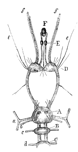
Diagram of the anterior portion of the nervous system in Lepas fascicularis. A. First thoracic or infra-œsophageal ganglion. B. Second thoracic. C. Third thoracic ganglion. D. Supra-œsophageal ganglion. E. The two ophthalmic ganglia. F. Double eye. a. Nerve going to first cirrus; b, to the muscles below the first cirrus; c, to the second cirrus; d, to the third; e, nerves running to the ovaria; f, double nerves supplying the sack and peduncle.
Ophthalmic Ganglia and Eyes.—Owing to Professor Leidy’s[16] discovery of eyes in a Balanus, I was led to look for them in the Lepadidæ. Extending from the front of the two supra-œsophageal ganglia, two chords may be seen in Lepas fascicularis (of which a rude diagram is here given), to run into two small, perfectly distinct oval ganglia (E), which are not united by any transverse commissure. From the opposite ends of these two ganglia smaller nerves run, and, bending inwards at right angles, enter, beyond the middle, an elongated (F), almost black, eye, composed of two eyes united together. Although in outline the eye appears single, two lenses can be distinctly seen at the end, directed upwards and towards the ganglia; two pigment-capsules can also be distinguished; these are deep and cup-formed, and of a dark reddish-purple. The following measurements will show the proportions of the parts in a specimen of the Lepas fascicularis having a capitulum 4/10ths of an inch in length.
Double eyelength26/6000
width13/6000
Diameter of single lens 6/6000
Ophthalmic ganglionlength16/6000
breadth11/6000
Supra-œsophageal ganglion, transverse or longest axis of both together126/6000
Supra-œsophageal ganglion, longitudinal axis of45/6000
Infra-œsophageal ganglion, transverse axis of120/6000
Infra-œsophageal ganglion, longitudinal axis of114/6000
In Conchoderma aurita the ophthalmic ganglia are much smaller, and nearer to the supra-œsophageal ganglion, than in L. fascicularis. In Alepas cornuta the ophthalmic chords run towards each other from the two distant and separate supra-œsophageal ganglia; and the ophthalmic ganglia, (instead of being quite separate, as in L. fascicularis,) are united by their front ends, and the two eyes instead of standing some way in front, with nerves running to them, are embedded on the double ophthalmic ganglion; the pigment-capsules here, also, have the shape of mere saucers, and are joined back to back, with the two lenses projecting far out of them. In neither sex of Ibla could I perceive that the eye was double. In Pollicipes spinosus the ophthalmic ganglion stands in front of the single supra-œsophageal ganglion, and shows no signs of being formed of a lateral pair; the eyes themselves, however, differently from, in all the foregoing cases, are, though approximate, quite distinct. In Pollicipes mitella I did not see the eyes; but the ophthalmic ganglion consists, as I believe, of a single globular one, placed exactly between the two globular, supra-œsophageal ganglia, all three being of nearly equal size. Professor Leidy does not mention the ophthalmic ganglia; hence I infer that in Balanus, which is a more highly organised Cirripede, they are fused into the supra-œsophageal ganglion.
In all the genera, the double eye is seated deep within the body; it is attached by fibrous tissue to the radiating muscles of the lowest part of the œsophagus, and lies actually on the upper part of the stomach; consequently, a ray of light, to reach the eye, has to pass through the exterior membrane and underlying corium connecting the two scuta, and to penetrate deeply into the body. In living sessile Cirripedes, vision seems confined to the perception of the shadow of an object passing between them and the light; they instantly perceived a hand passed quickly at the distance of several feet between a candle and the basin in which they were placed.
As the infra-œsophageal ganglion sends nerves to the trophi and to the first pair of cirri, it must correspond to the segments, from the fourth to the ninth inclusive, of the archetype crustacean. The state of the supra-œsophageal and ophthalmic ganglia appears to me very interesting: I do not believe that in any mature ordinary crustacean, the first or ophthalmic ganglion can be shown to be distinct from the two succeeding ganglia, or to be itself composed of a pair laterally distinct. The ganglia, corresponding with the second and third segments of the body, which should normally support two pair of antennæ, are in the Lepadidæ united together; but laterally they are generally distinct in outline, and are actually separate in Alepas: the supra-œsophageal ganglion shows also its double nature, by giving rise to a pair of large double nerves, evidently corresponding with the two pair of antennular nerves in ordinary crustaceans. The embryonic condition of the whole supra-œsophageal portion of the nervous system in the Lepadidæ, corresponds with the rudimentary state of the only organ of sense supplied by it, namely, the eye, which in size and general appearance has retrograded to the state in which it was in, during the first stage of development of the larva;—I have used the term embryonic, because, in the embryos of ordinary crustacea, all the ganglia are at first longitudinally distinct, and laterally quite separate. The conclusion at which we before arrived from studying the metamorphoses, namely, that the whole peduncle and capitulum consisted of the first three segments of the head, is beautifully supported by the structure of the nervous system, in which these parts are seen to be supplied with nerves exclusively from the supra-œsophageal ganglion: now in ordinary crustacea the supra-œsophageal ganglion sends nerves to the eyes and the two pair of antennæ corresponding, as is known by embryological dissections, to the first three segments of the body. Moreover, it is asserted that the carapace which covers the thorax in crustacea, is not formed by the development of the first segment; and this, likewise, may be inferred to be the case with the peduncle and capitulum in the Lepadidæ, as the nerves of the ophthalmic ganglia go exclusively to the eyes. Finally, I may remark that in Pollicipes, looking to the whole nervous system, the state of concentration nearly equals that in certain macrourous decapod crustaceans, for instance the Astacus marinus, of which a figure is given by Milne Edwards.
Olfactory Organs.—In the outer maxillæ, at their bases where united together, but above the basal fold separating the mouth from the body, there are, in all the genera, a pair of orifices (Pl. X, fig. 16); these are sometimes seated on a slight prominence, as in Lithotrya, or on the summit of flattened tubes (Pl. X, fig. 17), projecting upwards and towards each other, as in Ibla, Scalpellum, and Pollicipes. In Ibla these tubular projections rise from almost between the outer and inner maxillæ. It is impossible to behold these organs, and doubt that they are of high functional importance to the animal. The orifice leads into a deep sack lined by pulpy corium, and closed at the bottom. The outer integument is inflected inwards, (hence periodically moulted,) and becoming of excessive tenuity, runs to near the bottom of the sack, where it ends in an open tube: so excessively thin is this inflected membrane, that, until examining Anelasma, I was not quite certain that I was right in believing that the outer integument did not extend over the whole bottom. I several times saw a nerve of considerable size entering and blending into a pulpy layer at the bottom of the sack of corium; but I failed in tracing to which of the three pair of nerves, springing from the front end of the infra-œsophageal ganglion, it joined. I can hardly avoid concluding, that this closed sack, with its naked bottom, is an organ of sense; and, considering that the outer maxillæ serve to carry the prey entangled by the cirri towards the maxillæ and mandibles, the position seems so admirably adapted for an olfactory organ, whereby the animal could at once perceive the nature of any floating object thus caught, that I have ventured provisionally to designate the two orifices and sacks as olfactory.
Acoustic (?) Organs.—A little way beneath the basal articulation of the first cirrus (Pl. IX, fig. 4 d, and Pl. IV, fig. 2 e), on each side, there may be seen a slight swelling, and on the under side of this, a transverse slit-like orifice, 1/20th of an inch in length in Conchoderma, but often only half that size. In Ibla this orifice is seated lower down (Pl. IV, fig. 8 a´, e), between the bases of the first and second cirri, which are here far apart: in Alepas cornuta it is placed rather nearer to the adductor scutorum muscle, namely, beneath the mandibles. The orifice leads into a rather deep and wide meatus; the external integument is turned in for a short distance, widening a little, and then ends abruptly. The meatus, enlarging upwards, is lined by thick pulpy corium, and is closed at the upper end; from its summit is suspended a flattened sack of singular and different shapes in the different genera. This, the so-called acoustic sack of Conchoderma virgata, is figured Pl. IX, fig. 6. The deep and wide notch faces towards the posterior end of the animal; the inferior lobe, thus almost cut off, is flattened in a different plane from the upper part; the lobe is lodged in a little pouch of corresponding form, leading from the open meatus in which the upper part is included. In Conchoderma aurita, the top of the acoustic sack is narrower and more constricted, the whole more rounded, and the lobe more turned down. In Lepas fascicularis the notch is not so deep or wide, and the lobe larger. In Ibla Cumingii the sack is of the shape of a vase, with one corner folded over. In Scalpellum vulgare it is small, oval, with the lower end much pushed in, and furnished with a little crest. Lastly, in Pollicipes mitella it is simply oval. In all cases the sack is empty, or contains only a little pulpy matter: it consists of brownish, thick, and remarkably elastic tissue, formed, apparently, of transverse little pillars, becoming fibrous on the outside, and with their inner ends appearing like hyaline points. The mouth of the acoustic sack (removed in the drawing) is closed by a tender diaphragm, through which I saw what I believe was a moderately-sized nerve enter; I have not yet succeeded in tracing this nerve. The first pair of cirri seem, to a certain extent, to serve as antennæ, and therefore the position of an acoustic organ at their bases, is analogous to what takes place in crustacea; but there are not here any otolites, or the siliceous particles and hairs, as described by Dr. Farre, in that class. Nevertheless, the sack is so highly elastic, and its suspension in a meatus freely open to the water, seems so well adapted for an acoustic organ, that I have provisionally thus called it. In the larva, as I have shown, a pouch, certainly serving for some sense, I believe for hearing, is seated in quite a different position at the anterior end of the carapace. I may mention that I found sessile Cirripedes very sensitive of vibrations in objects adjoining them, though not, apparently, of noises in the air or water. In a group of specimens, I could not touch one even most delicately with a needle, without all the adjoining ones instantly withdrawing their cirri; it made no difference if the one touched had its operculum already closed and motionless.
Reproductive System,—Male Organs.—All the Cirripedia which I have hitherto examined, with the exception of certain species of Ibla and Scalpellum, are hermaphrodite or bisexual.[17] I shall so fully describe the sexual relations of the several species of these two genera, under their respective headings, and at the end of the genus of Scalpellum, that I will not here give even an abstract of the grounds on which my firm belief is based, that the masculine power of certain hermaphrodite species of Ibla and Scalpellum, is rendered more efficient by certain parasitic males, which, from their not pairing, as in all hitherto known cases, with females, but with hermaphrodites, I have designated Complemental Males.
The male organs have been well described by M. Martin St. Ange, whose observations have since been confirmed by R. Wagner.[18] The testes are small, often leaden-coloured, either pear or finger-shaped, or branched like club-moss,—these several forms sometimes occurring in the same individual; they coat the stomach, enter the pedicels, and even the basal segments of the rami of the cirri, and in some genera occupy certain swellings on the thorax and prosoma, and in others the filamentary appendages: the testes seen in the apex in one of these appendages in Conchoderma, is represented in Pl. IX, fig. 5. The two vesiculæ seminales are very large; they lie along the abdominal surface of the thorax, and generally (but not in some species of Scalpellum) enter the prosoma, where their broad ends are often reflexed; here the branched vessels leading from the testes enter. The membrane of the vesiculæ seminales is formed of circular fibres; and is, I presume, contractile, for I have seen the spermatozoa expelled with force from the cut end of a living specimen. The two canals leading from the vesiculæ generally unite in a single duct at the base of the penis; but in Conchoderma aurita, half-way up it. The probosciformed penis, except in certain species of Scalpellum, is very long; it is capable of the most varied movements; it is generally hairy, especially at the end; it is supported on a straight unarticulated basis, which in Ibla quadrivalvis alone (Pl. IV, fig. 9 a), is of considerable length; in this species, the upper part is seen to be as plainly articulated as one of the cirri; in Alepas, the articulations are somewhat less plain, and in the other genera, the organ can be said only to be finely ringed, but these rings no doubt are in fact obscure articulations. In the females of Ibla Cumingii and Scalpellum ornatum, there is, of course, no penis.